
Page 946 - Hematology_ Basic Principles and Practice ( PDFDrive )
P. 946
Chapter 56 Conventional and Molecular Cytogenomic Basis of Hematologic Malignancies 829
1 2 3 4 5
6 7 8 9 10 11 12
13 14 15 16 17 18
19 20 21 22 X Y
A
1 2 3 4 5
6 7 8 9 10 11 12
13 14 15 16 17 18
19 20 21 22 X Y
B
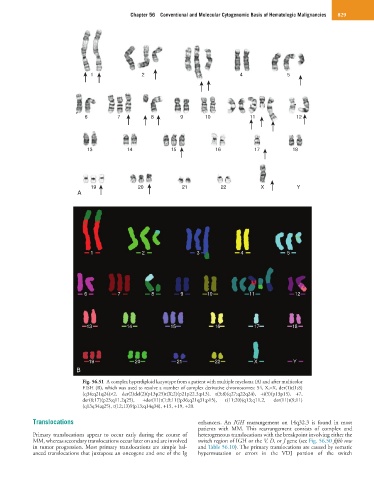
Fig. 56.51 A complex hyperdiploid karyotype from a patient with multiple myeloma (A) and after multicolor
FISH (B), which was used to resolve a number of complex derivative chromosomes: 54, X,−X, der(1)t(1;8)
(q34;q21q24)×2, der(2)del(2)(p13p25)t(X;2)(p21p22.3;p13), t(3;8)(q27;q22q24), +i(5)(p13p15), +7,
der(8;17)(p23;q11.2q25), +der(11)t(1;9;11(?p36;q21q31;p15), t(11;20)(q13;q11.2, der(11)t(9;11)
(q13q34;q25), t(12;13)9(p13;q14q34), +15, +19, +20.
Translocations enhancers. An IGH rearrangement on 14q32.3 is found in most
patients with MM. This rearrangement consists of complex and
Primary translocations appear to occur early during the course of heterogeneous translocations with the breakpoint involving either the
MM, whereas secondary translocations occur later on and are involved switch region of IGH or the V, D, or J gene (see Fig. 56.50 fifth row
in tumor progression. Most primary translocations are simple bal- and Table 56.10). The primary translocations are caused by somatic
anced translocations that juxtapose an oncogene and one of the Ig hypermutation or errors in the VDJ portion of the switch