
Page 954 - Hematology_ Basic Principles and Practice ( PDFDrive )
P. 954
Chapter 56 Conventional and Molecular Cytogenomic Basis of Hematologic Malignancies 837
MYC Amplification a sequence-specific transcriptional repressor that is upregulated; 25%
of ABC-DLBCL have deletions of PRDM1 as a result of truncated
mutations, or genomic deletions, while an additional fraction of
patients lacks the PRDM1 protein as a result of transcriptional repres-
sion by a constitutively active translocated BCL6 gene.
Patients with non-BL may have MYC translocations without
additional mutation in BCL2 or BCL6 and they are described as
having single hit lymphomas. Although the most recent studies sug-
gested that patients with MYC+, but BCL2− and BCL6− DLBCL do
not have an inferior outcome when compared to those who are MYC
negative, the clinical relevance of MYC rearrangement as sole abnor-
mality in DLBCL is still controversial with some studies indicating
an adverse prognosis.
Double hit lymphoma (DHL) refers to B-cell lymphoma with
multiple activating oncogenes, one of them being MYC. Among
DHL, those with alterations in MYC and BCL2 are the most common
by far, occurring in 87% of patients, followed by MYC and BCL6
rearrangements, found in 5% of patients. Triple hit lymphoma
(THL) involves rearrangements of MYC, BCL6, and BCL2 in a single
A cell and are observed in about 8% of patients with DLBCL
(Fig. 56.58).
DHL can be detected cytogenetically but cryptic gene rearrange-
ments may be missed. FISH therefore should be used for the detection
of DHL and THL. FISH studies can be successfully performed using
formalin-fixed paraffin embedded tissue sections as well as fresh
smears, touch-cell imprints.
Because one of the translocations may be cryptic, a combination
of three FISH probes, labeled with three colors, is especially useful
in identifying these very complex translocations (see Fig. 56.43).
Non-IG genes also act as MYC translocation partners in 35% to 53%
of MYC rearranged DLBCL. The t(8;9)(q24;p13), which results in
the juxtaposition of MYC to PAX5, has been frequently reported,
accounting for 20% of non-IG/MYC rearrangement. Both the DHL
and THL have complex karyotypes. Complex karyotypes are not seen
in BL, which by definition always have single abnormality.
The detection of increased number of cells expressing MYCs
B correlates well with the presence of gene rearrangements, but the
relationship varies. Tumors with more than 70% of MYC-positive
cells usually are associated with gene rearrangements, but MYC
translocations can be present in up to 17% of cases with less than
30% MYC-positive cells. The variability between the protein expres-
sion and gene alterations makes it difficult to recommend the use of
MYC immunohistochemistry as a screening method to detect gene
rearrangements. The current recommendation is that all patients with
DLBCL have FISH for MYC rearrangements because combined
alterations in MYC, BCL2, and BCL6 confers a more aggressive
disease phenotype.
All patients with MCL exhibit t(11;14)(q13;q32) abnormality as
detected by FISH (see Fig. 56.55 top panel). t(11;14)(q13;q32) is also
found in a variety of other B-cell malignancies, including MM,
splenic lymphoma with villous lymphocytes, and B-cell prolympho-
cytic leukemia. Most breakpoints on chromosome 11, band q13, are
dispersed over a region of approximately 130 kb centromeric to the
cyclin D1 (CCND1) gene. At the molecular level, the BCL1 locus
C (CCND1) on chromosome 11q13 is juxtaposed to an enhancer
sequence within the IGH gene on 14q32, leading to overexpression
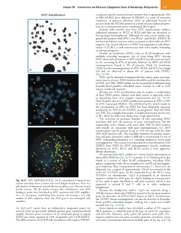
Fig. 56.57 MYC AMPLIFICATION. (A) A conventional G-banded bone of the cyclin D gene (involved in cell cycle control), which is not
marrow metaphase from a patient with non-Hodgkin lymphoma. Note very expressed in normal B and T cells or in other malignant
pale doublets of chromatin material all over metaphase cells. They are termed lymphomas.
double minutes. (B) All double minutes after hybridization with MYC Because the breakpoints within 11q13 are scattered along a
breakapart probe were hybridized to MYC consistent with MYC amplifica- 130-kb distance, dual-color FISH has proved to be the most sensitive
tion. (C) Interphase, nondividing bone marrow cell showing a very high assay for detection of IGH-CCND1 fusion in MCL. Furthermore,
number of MYC amplicon. Note that MYC gene is not rearranged, only the CCND1 fusion rearrangement can also be detected in formalin-
amplified. fixed, paraffin-embedded samples, making this a rapid and reliable
method (see Fig. 56.42A).
the 3p11–p12 region have an independent prognostic power for Gene expression studies have identified a subset of D1-negative
survival based on previously defined optimal gene expression–based MCL patients, so called because of the lack of cyclin D1 expression
models. Somatic point mutations of the polycomb-group oncogene and t(11;14). However, both cyclin D1–positive and cyclin D1–
EZH2 have been reported in 22% of patients with GCB-DLBCL. negative patients have the same secondary genomic alterations. These
The differentiation of GCB B cells into plasma cells requires PRDM1, secondary chromosomal alterations include gains of 3q, 8q, and 15q