
Page 960 - Hematology_ Basic Principles and Practice ( PDFDrive )
P. 960
Chapter 56 Conventional and Molecular Cytogenomic Basis of Hematologic Malignancies 843
(q11q32.1), t(14;14)(q11;q32.1), and t(7;14)(q35;q32.1) (see Fig.
56.60). In patients showing these karyotypic changes, TCL1 is found
to be dysregulated.
Adult T-cell leukemia/lymphoma is associated with human T-cell
lymphotropic virus type 1. The most frequent genetic lesions include
altered expression of CDKN2 (cyclin-dependent kinase inhibitor)
gene on 9p21 (15%–20%) and loss of heterozygosity at 6q15 to q21.
The most frequent chromosomal abnormalities are gain of 3p, 7q,
and 14q and loss of 6q and 13q. Translocations involving 14q32 or
14q11 are frequently observed.
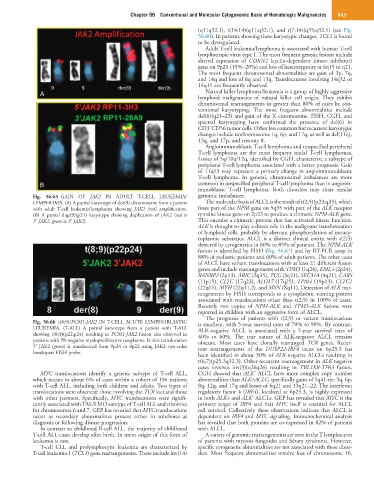
9 9 der(9) der(9)
A Natural killer lymphoma/leukemia is a group of highly aggressive
lymphoid malignancies of natural killer cell origin. They exhibit
chromosomal rearrangements in greater than 80% of cases by con-
ventional karyotyping. The most frequent abnormalities include
del(6)(q21–23) and gain of the X chromosome. FISH, CGH, and
spectral karyotyping have confirmed the presence of del(6) in
−
CD3 CD56 tumor cells. Other less common but recurrent karyotypic
changes include isochromosome 1q, 6p, and 17q, as well as del(11q),
13q, and 17p, and trisomy 8.
Angioimmunoblastic T-cell lymphoma and unspecified peripheral
T-cell lymphoma are the most frequent nodal T-cell lymphomas.
Losses of 5q/10q/12q, identified by CGH, characterize a subtype of
peripheral T-cell lymphoma associated with a better prognosis. Gain
of 11q13 may represent a primary change in angioimmunoblastic
T-cell lymphoma. In general, chromosomal imbalances are more
B common in unspecified peripheral T-cell lymphoma than in angioim-
munoblastic T-cell lymphoma. Both disorders may share similar
Fig. 56.65 GAIN OF JAK2 IN ADULT T-CELL LEUKEMIA/ genomic imbalances.
LYMPHOMA. (A) A partial karyotype of der(9) chromosome from a patient The molecular basis of ALCL is the result of t(2;5)(p23;q35), which
with adult T-cell leukemia/lymphoma showing JAK2 (red) amplification. fuses part of the NPM gene on 5q35 with part of the ALK receptor
(B) A partial dup(9)(p24) karyotype showing duplication of JAK2 (red is tyrosine kinase gene on 2p23 to produce a chimeric NPM-ALK gene.
5′ JAK2, green is 3′ JAK2). This encodes a chimeric protein that has activated kinase function.
ALK is thought to play a direct role in the malignant transformation
of lymphoid cells, probably by aberrant phosphorylation of intracy-
toplasmic substrates. ALCL is a distinct clinical entity, with t(2;5)
detected by cytogenetics in 60% to 85% of patients. The NPM-ALK
fusion is identified by FISH (Fig. 56.67) and by RT-PCR assay in
88% of pediatric patients and 60% of adult patients. The other cases
of ALCL have variant translocations with at least 21 different fusion
genes and include rearrangements with TPM3 (1q24), EML4 (2p24),
RANBP2 (2q13), ARIC (2q35), TCG (3q21), SEC31A (4q21), CARS
(11p15), CLTC (17q23), ALO17 (17q25), TPM4 (19p13), CLTC1
(22q11), MYH (22q11.2), and MSN (Xq11). Detection of ALK rear-
rangements by FISH corresponds to a cytoplasmic staining pattern
associated with translocations other than t(2;5) in 100% of cases.
Recently two copies of NPM-ALK and TPM3-ALK fusions were
reported in children with an aggressive form of ALCL.
The prognosis of patients with t(2;5) or variant translocations
Fig. 56.66 t(8;9)/PCM1-JAK2 IN T-CELL ACUTE LYMPHOBLASTIC is excellent, with 5-year survival rates of 70% to 90%. By contrast,
LEUKEMIA. (T-ALL) A partial karyotype from a patient with T-ALL ALK-negative ALCL is associated with a 5-year survival rates of
showing t(8;9)(p22;p24) resulting in PCM1-JAK2 fusion also observed in 40% to 60%. The true nature of ALK-negative ALCL remains
patients with Ph-negative myeloproliferative neoplasms. In this translocation obscure. Most cases have clonally rearranged TCR genes. Recur-
5′ JAK2 (green) is translocated from 9p24 to 8p22 using JAK2 two-color rent rearrangements of the DUSP22-IRF4 locus on 6p25.3 has
breakapart FISH probe. been identified in about 30% of ALK-negative ALCLs resulting in
t(6;7)(p25.3q32.3). Other recurrent rearrangement in ALK negative
cases involves inv(3)(q26q26) resulting in TBL1XR-TP63 fusion.
−
MYC translocations identify a genetic subtype of T-cell ALL, CGH showed that ALK ALCL have more complex copy number
which occurs in about 6% of cases within a cohort of 196 patients abnormalities than ALK+ACLC, specifically gains of 1q41-ter, 5q, 6p,
with T-cell ALL, including both children and adults. Two types of 8q, 12q, and 17q and losses of 6q21 and 13q21–22. The interferon
translocations were observed: those involving the TCR loci and those regulatory factor 4 (IRF4), localized at 6p25.3, is highly expressed
−
with other partners. Specifically, MYC translocations were signifi- in both ALK+ and ALK ALCLs. GEP has revealed that MYC is the
cantly associated with TAL/LMO subtype of T-cell ALL and trisomies primary target of IRF4 and that MYC itself is essential for ALCL
for chromosomes 6 and 7. GEP has revealed that MYC translocations cell survival. Collectively these observations indicate that ALCL is
occur as secondary abnormalities present either in subclones at dependent on IRF4 and MYC signaling. Immunochemical analysis
diagnosis or following disease progression. has revealed that both proteins are co-expressed in 82% of patients
In contrast to childhood B-cell ALL, the majority of childhood with ALCL.
T-cell ALL cases develop after birth. In utero origin of this form of A variety of genomic rearrangements are seen in the T lymphocytes
leukemia is rare. of patients with mycosis fungoides and Sézary syndrome. However,
T-cell CLL and prolymphocytic leukemia are characterized by specific cytogenetic abnormalities are not associated with these disor-
T-cell leukemia 1 (TCL1) gene rearrangements. These include inv(14) ders. Most frequent abnormalities involve loss of chromosome 10,