
Page 964 - Hematology_ Basic Principles and Practice ( PDFDrive )
P. 964
Chapter 56 Conventional and Molecular Cytogenomic Basis of Hematologic Malignancies 847
ALL in recipient cells
1 2 3 4 5
B
6 7 8 9 10 11 12
13 14 15 16 17 18
19 20 21 22 X Y
A C
MDS in donor cells
del EGR1(red) del p53(red)
del 7q31 (red)
1 2 3 4 5
6 7 8 9 10 11 12
E disomy 21 (red), X (green), Y (aqua)
13 14 15 16 17 18
19 20 21 22 X Y
D F
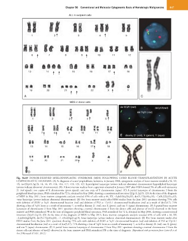
Fig. 56.69 DONOR-DERIVED MYELODYSPLASTIC SYNDROME (MDS) FOLLOWING CORD BLOOD TRANSPLANTATION IN ACUTE
LYMPHOBLASTIC LEUKEMIA. (A) At diagnosis of acute lymphoblastic leukemia, in January 2006, cytogenetic analysis of bone marrow revealed a 56, XY,
+X, inv(2)(p11.2q13), +4, +6, +9, +14, +14, +17, +18, +21, +21 hyperdiploid karyotype (arrows indicate abnormal chromosomes) hyperdiploid karyotype
(arrows indicate abnormal chromosomes). (B). A bone marrow nucleus from a specimen obtained in January 2007 after FISH showed 5% of cells with tetrasomy
21 (red signals), two copies of X chromosome (green signals), and one copy of Y chromosome (aqua). (C) A partial karyotype of chromosome 2 from the
peripheral blood specimen, PHA-stimulated for 72 h, obtained in May 2006, showing a constitutional inversion (2)(p11.2q13). (D) At the time of the diagnosis
of MDS in May 2011, bone marrow cytogenetic analysis revealed 65% of cells with a 44, XY, −3,del(4)(q23q33), der(5;17)(p10;q10), −7,t(8;22)(p21;q13),
+mar karyotype (arrows indicate abnormal chromosomes). (E) Five bone marrow nuclei after FISH studies from the June 2011 specimen showing 75% cells
with deletion of EGR1 at 5q31 chromosomal location (red) and deletion of P53 at 17p13.1 chromosomal localization (red) as a result of der(5;17); 71%
showing a loss of 7q31 locus as a result of monosomy 7, as well as disomy 21 (red), one X (green), and one Y (aqua) chromosome. (F) A partial bone marrow
karyotype of chromosome 2 from May 2011 specimen showing a normal chromosome 2 from the donor cells and absence of inv(2) observed in the bone
marrow and PHA-stimulated PB at the time of diagnosis peripheral blood specimen, PHA stimulated for 72 h, obtained in May 2006, showing a constitutional
inversion (2)(p11.2q13). (D) At the time of the diagnosis of MDS in May 2011, bone marrow cytogenetic analysis revealed 65% of cells with a 44, XY,
−3,del(4)(q23q33), der(5;17)(p10;q10), −7, t(8;22)(p21;q13), 1mar karyotype (arrows indicate abnormal chromosomes). (E) Five bone marrow nuclei after
FISH studies from the June 2011 specimen showing 75% cells with deletion of EGR1 at 5q31 chromosomal location (red) and deletion of P53 at 17p13.1
chromosomal localization (red) as a result of der(5;17); 71% showing a loss of 7q31 locus as a result of monosomy 7, as well as disomy 21 (red), one X (green),
and one Y (aqua) chromosome. (F) A partial bone marrow karyotype of chromosome 2 from May 2011 specimen showing a normal chromosome 2 from the
donor cells and absence of inv(2) observed in the bone marrow and PHA-stimulated PB at the time of diagnosis. (Reproduced with permission from Cotter R et al:
Am J Hematol 87:931, 2012.)