
Page 1766 - Williams Hematology ( PDFDrive )
P. 1766
1740 Part XI: Malignant Lymphoid Diseases Chapter 107: Myeloma 1741
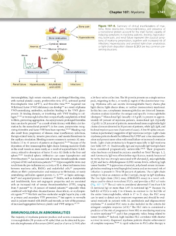
Bone pain Figure 107–6. Summary of clinical manifestations of mye-
loma. Bone destruction, immunodeficiency, and presence of
a monoclonal protein account for the main factors capable of
inducing symptoms in myeloma patients. Anemia, hypercalce-
Bone destruction Hypercalcemia mia, bone pain, and renal failure represent the classical symp-
toms of myeloma presentation, together with increased risk of
Cytokine release infections. Hyperviscosity and amyloid light-chain amyloidosis
Anemia or light-chain deposition disease (LCDD) are less common pre-
Marrow infiltration senting situations.
Albumin
M-spike
Myeloma
Monoclonal protein Immunodeficiency
Renal failure Hyperviscosity Amyloidosis Infections
and LCDD
immunoglobins, high serum viscosity, and a prolonged bleeding time, a 24-hour urine collection. The M-protein presents as a single narrow
with normal platelet counts, prothrombin time (PT), activated partial peak, migrating in the γ, or rarely β, region of the densitometer trac-
thromboplastin time (aPTT), and thrombin time. 248,249 Acquired von ing. Myeloma cells can secrete immunoglobulin heavy chains plus
Willebrand factor (VWF) deficiency can develop as a result of plasma light chains, light chains alone, or neither (nonsecretory myeloma).
250
VWF-neutralizing antibodies, antibodies binding to the VWF glyco- In this last case, cytoplasmic immunoglobulins are detected. Immun-
protein 1b binding domain, or interfering with VWF binding to col- ofixation analysis identifies the unique and specific immunoglobulin
lagen, 251,252 or immunoglobulins that nonspecifically coat platelets or bind idiotypes. Monoclonal IgG (usually >3.5 g/dL) is present in approx-
47
to fibrin, preventing aggregation. An asymptomatic prolonged thrombin imately 60 percent of myeloma patients, monoclonal IgA (typically
time can also be present, as a result of interference with fibrin clot for- >2 g/dL) in 20 percent of patients, monoclonal immunoglobulin light
253
mation by the monoclonal protein ; in few cases, paraproteins recog- chains alone are detected in 20 percent of patients, while IgD, IgM, and
254
nizing thrombin and factor VIII have been reported. 253,255 Bleeding may biclonal myeloma are rare (5 percent of cases). A low M-spike concen-
also result from progression of disease, renal insufficiency, infections, tration is particularly suggestive of IgD myeloma isotype. Light-chain
therapy-related toxicity, invasive procedures, and anoxia/thrombosis in myeloma patients should be followed by UPEP and urine immunofix-
the capillary circulation. Bleeding is more common in systemic AL amy- ation and present more often with renal failure or increased creatinine
loidosis (15 to 41 percent of patients at diagnosis), 256–258 because of the levels. Light-chain proteinuria is frequent especially in IgD myeloma
deposition of free immunoglobin light chains forming insoluble fibrils (see Table 107–3). Traditionally, IgA and especially IgD isotypes have
in the small vessels or, more rarely, as a result of acquired factor X defi- been considered prognostically unfavorable. 280,281 Their prognostic
ciency, related to absorption of factor X onto AL fibrils in the liver and value has been confirmed in patients enrolled on Total Therapy 1, 2,
spleen. 259,260 Amyloid splenic infiltration can cause hyposplenism and and 3 protocols; IgD was of borderline significance, mainly because of
thrombocytosis. An increased risk of venous thromboembolic events its rarity, but was strongly associated with elevated β -microglobulin
260
2
is typical of MG and myeloma patients. 261,262 Hypercoagulable states may (β M) and lactic dehydrogenase (LDH) serum levels, reflecting high
2
result from the pro-inflammatory activity of IL-6, abnormal interactions tumor burden. Suppression of normal, polyclonal serum immuno-
282
between myeloma cells, BMSCs and endothelial cells, paraprotein globulins resulting in total hypoglobulinemia and an increased risk of
263
effects on fibrin polymerization and resistance to fibrinolysis, or rarely infection is present in 70 to 90 percent of patients. The κ light-chain
neutralizing antibodies against protein C, S, 264,265 or lupus anticoagu- isotype is twice as common as the λ isotype, except in IgD myeloma.
lant and acquired protein C resistance. IMiDs, such as thalidomide The free light chain (FLC) assay (FREELITE assay) is a novel tech-
267
266
and lenalidomide, have anti-angiogenic properties and are associated nique used to detect monoclonal FLCs, and provides a FLC κ:λ ratio
with an increased risk of venous thromboembolism (VTE), ranging (Fig. 107–7). The κ:λ ratio is considered abnormal if less than 0.26
283
from 5 percent to 18 percent of treated patients, especially when (λ-restricted Ig) or more than 1.65 (κ-restricted Ig). Because the
284
269
268
combined with high-dose dexamethasone, doxorubicin, or erythropoi- half-life of FLCs is only 2 to 4 hours, in contrast to the half-life of
etic agents. 270–274 Warfarin and low-molecular-weight heparin play a role the entire immunoglobulin, which is 17 to 21 days, the FLC assay
in primary and secondary VTE prevention, but aspirin is normally can be used to detect early treatment responses and should be eval-
275
used in patients treated with IMiDs and steroids, in view of the presence uated routinely in patients with AL amyloidosis and oligosecretory
of increased aggregation between platelet and VWF antigens. 276–279 myeloma. A normal FLC ratio is also included in the criteria for
285
stringent complete response (sCR). The FLC ratio is considered a
8
predictor of the risk of progression from MG or smoldering myeloma
IMMUNOGLOBULIN ABNORMALITIES to active myeloma 286,287 and it has prognostic value, being related to
285
The majority of myeloma patients produce and secrete a monoclonal tumor burden. Indeed, high baseline FLC correlates with shorter
immunoglobulin (M-protein or M-spike) that can be detected by pro- survival in newly diagnosed myeloma patients despite achievement
tein electrophoresis of the serum (SPEP) and/or of urine (UPEP) after of complete response. 288,289 A rapid reduction in FLCs after therapy is
Kaushansky_chapter 107_p1733-1772.indd 1741 9/21/15 12:34 PM