
Page 1767 - Williams Hematology ( PDFDrive )
P. 1767
1742 Part XI: Malignant Lymphoid Diseases Chapter 107: Myeloma 1743
Normal immunoglobulin Free light chains Figure 107–7. Free light-chain assay description. Normal
Ig variable regions immunoglobulins are composed of two heavy chains and two
light chains, which together form a constant region and a variable
region, capable of recognizing specific antigens. The free light-
chain assay is used to quantify the amount of free light chains in
Ig constant regions myeloma patients and to specifically recognize a “hidden” anti-
genic region (in red) that is normally not detectable from intact
Heavy immunoglobulins.
chain
Hidden region
Previously hidden
region
Light chain
also linked to inferior overall and event-free survival, suggesting the plasma cells can be indistinguishable from normal plasma cells, char-
presence of highly proliferative myeloma cells, particularly sensitive acterized by abundant basophilic cytoplasm and round, eccentrically
to combination chemotherapy. 290 located nuclei, with “clock-face” or “spoke-wheel” chromatin without
nucleoli, or by bizarre plasma cells with giant cellular size, open chro-
matin, and punched nucleoli (indicating increased transcriptional
MARROW FINDINGS activity), a high frequency of binucleate or multinucleate cells, and the
A marrow aspirate or biopsy showing plasmacytosis is a key compo- presence of inclusion globules of condensed or crystallized cytoplasmic
nent for the diagnosis of myeloma. The marrow can be evenly infiltrated immunoglobulin, including Russell bodies (cherry-red refractive round
(diffuse involvement) but more commonly displays considerable site- bodies), multiple pale bluish-white, grape-like accumulations (Mott
to-site variability (focal involvement). The percentage of plasma cells cells or Morula cells), crystalline rods, glycogen-rich IgA (flame cells),
291
can range from 10 percent to a virtual complete replacement of marrow. or other inclusions. These abnormal cells are characteristic of plasmab-
Occasionally, the biopsy specimen may contain a normal proportion lastic myeloma, 292,293 a poor prognostic type of myeloma with a high
294
of plasma cells as a result of the patchy marrow involvement. Myeloma number of mitotic figures (Figs. 107–8 to 107–10). Myeloma cells are
diagnosis is also considered in the presence of less than 10 percent of clonal by definition and produce either κ or λ light chains, which are
chain restricted plasma cells if symptoms are reported or after histo- present in the cytoplasm but not on the membrane surface. A κ:λ ratio
295
pathologic confirmation of an intraosseous or extraosseous plasmacy- greater than 4:1 (normal 2:1 ) or less than 1:2 is considered an index
toma. By light microscopy, the morphologic appearance of myeloma of κ or λ monoclonality, distinguishing this condition from reactive
A B
C
Mature
A B
Hof
Immature
C D
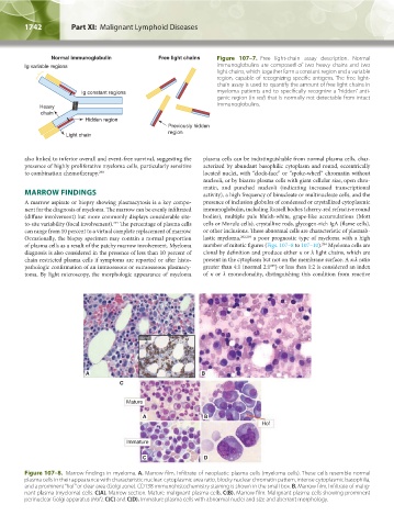
Figure 107–8. Marrow findings in myeloma. A. Marrow film. Infiltrate of neoplastic plasma cells (myeloma cells). These cells resemble normal
plasma cells in their appearance with characteristic nuclear: cytoplasmic area ratio, blocky nuclear chromatin pattern, intense cytoplasmic basophilia,
and a prominent “hof” or clear area (Golgi zone). CD138 immunohistochemistry staining is shown in the small box. B. Marrow film. Infiltrate of malig-
nant plasma (myeloma) cells. C(A). Marrow section. Mature malignant plasma cells. C(B). Marrow film. Malignant plasma cells showing prominent
perinuclear Golgi apparatus (Hof). C(C) and C(D). Immature plasma cells with abnormal nuclei and size and aberrant morphology.
Kaushansky_chapter 107_p1733-1772.indd 1742 9/21/15 12:34 PM