
Page 1769 - Williams Hematology ( PDFDrive )
P. 1769
1744 Part XI: Malignant Lymphoid Diseases Chapter 107: Myeloma 1745
Figure 107–11. Kappa-lambda staining.
plasmacytosis. Two-parameter flow cytometry staining for nuclear
296
DNA content by propidium iodide and anti-κ and anti-λ light-chains
can also be used to quantitate marrow involvement (Fig. 107–11).
297
Myeloma cells are normally CD138+, CD45–, CD38+, and CD19– ,
298
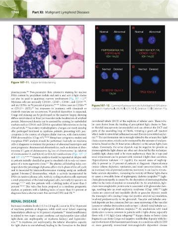
and are CD56+ in 70 percent of patients. 299–301 A few cases are CD20+ Figure 107–12. Common fluorescence in situ hybridization (FISH) abnor-
302
or CD117+ (KIT), but responses to treatment with rituximab or malities in myeloma. A. t(4;14). B. t(11;14). C. Deletion 13. D. Deletion 17p.
303
imatinib mesylate are uncommon. If amyloid deposition is suspected,
Congo red staining can be performed on the marrow biopsy, showing
diffuse involvement or focal perivascular niche localization of amyloid
protein. Microvessel density can be assessed by staining for endothelial convoluted tubule (DCT) of the nephron of tubular casts. These tubu-
markers such as CD131 and CD34 in specialized laboratories or during lar casts derive from the binding of precipitated light chains to Tam-
clinical trials. Secondary myelodysplastic changes can rarely develop m-Horsfall mucoprotein (uromodulin) and can obstruct the DCT and
304
after prolonged treatment in myeloma patients, presenting with pan- parts of the ascending loop of Henle, initiating a giant cell reaction
cytopenia in the context of a hypercellular marrow, with characteristic which leads to interstitial inflammation and fibrosis (interstitial nephri-
321
FISH abnormalities (Chap. 87). 305,306 Metaphase cytogenetic studies and tis). The cast formation rate is strongly related to the urinary free-light
interphase FISH analysis should be performed routinely on myeloma chain concentration, which can be estimated by the amount of total pro-
cells at diagnosis to evaluate the presence of abnormal karyotypes and teinuria, based on the 24-hour urine collection or the serum light chain
poor prognostic chromosomal abnormalities, such as deletion of chro- values. Conversely, the urine dipstick may be negative for protein as
mosome 17, gain of chromosome 1q, loss of chromosome 1p, deletion immunoglobulin light chains are often not detected by this technique.
of chromosome 13, and t(4;14) or t(14;16) translocations (Figs. 107–12 Lambda light chains tend to be more nephrotoxic than the κ type and
and 107–13). 63,307–310 Genetic analysis should be repeated at relapse only renal impairment can be present with minimal λ light-chain secretion.
in patients initially classified as genetic standard-risk to rule out emer- Hypercalcemia (calcium >11 mg/dL), the second cause of nephrop-
gence of a more aggressive clone. The plasma cell labeling index cor- athy, is present in 15 percent of patients at diagnosis. Hypercalcemia
311
responds to the percentage of plasma cells in the S-phase of the cell cycle creates volume depletion, natriuresis, and renal vasoconstriction, with
and is measured by immunofluorescence staining using an antibody an increased risk of prerenal azotemia; moreover, it can lead to intratu-
against 5-bromo-2′-deoxyuridine, which is actively incorporated by bular calcium deposition, increasing the toxicity of filtered light chains
322
DNA on marrow plasma cells. Actively cycling myeloma cells represent or cause a reversible form of nephrogenic diabetes insipidus. Light-
a small proportion of the total malignant cells, normally 0.5 percent on chain glomerulopathy is caused by the deposition of immunoglobulins
average, 312–317 with few patients with a labeling index of more than 5 either in the form of amyloid or nonamyloid. In AL amyloidosis, light-
percent. 318,319 This value has been proposed as a myeloma prognostic chain immunoglobulin proteinuria is associated with glomerular dam-
marker, as patients with a labeling index of more than 0.5 percent at age, resulting into an overt nephrotic syndrome (Chap. 108). Light
323
diagnosis have a shorter event-free survival (EFS) and OS. 318 chains are converted into insoluble fibrils or granular deposits inside
the mesangial cells causing Congo red-positive amyloid accumulation,
localized predominantly in the glomeruli. Vascular and tubular amy-
RENAL DISEASE loid deposits are less common, but can cause narrowing of the vascular
Increased creatinine levels (>1.5 to 2.0 mg/dL) occur in 30 to 50 percent lumens or tubular dysfunction such as type 1 (distal) renal tubular aci-
of myeloma patients at diagnosis, while overt renal failure requiring dosis or nephrogenic diabetes insipidus. AL amyloidosis with renal
324
hemodialysis affects up to 10 percent of patients. Renal insufficiency dysfunction is more common in patients with λ light-chains, especially
247
325
is related to two major causes: myeloma cast nephropathy (also called those with λ VI light-chain subgroup. Kappa chains or heavy-chain
light-chain cast nephropathy or myeloma kidney) and hypercalce- fragments can form Congo red-negative nonfibrillar deposits with lin-
320
mia. In myeloma cast nephropathy, the tubular absorptive capacity ear involvement of the basement membrane, in a condition called LCDD
for light chains is overwhelmed, leading to the formation in the distal or, more generally, monoclonal immunoglobulin deposition disease
Kaushansky_chapter 107_p1733-1772.indd 1744 9/21/15 12:34 PM