
Page 1773 - Williams Hematology ( PDFDrive )
P. 1773
1748 Part XI: Malignant Lymphoid Diseases Chapter 107: Myeloma 1749
TABLE 107-6. International Staging System
Stage I: β M <3.5
2
ALB ≥3.5
Stage II: β M <3.5
2
ALB <3.5
or
β M 3.5 to 5.5
2
Stage III: β M >5.5
2
ALB, serum albumin in g/dL; β M, serum β -microglobulin in mg/L. 231
2 2
Data from Greipp PR, San Miguel J, Durie BG, et al: International
staging system for multiple myeloma. J Clin Oncol 2005 May
20;23(15):3412–3420. A B C
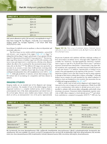
bone disease by skeletal survey in myeloma is observer-dependent and Figure 107–14. Plain x-rays of osteolytic lesions. Osteolytic lesions
potentially subjective. captured on plain x-rays or skeletal survey. A. Right humerus. B. Right
The ISS is based on two widely available parameters—serum β M femur. C. Right radius.
2
and albumin—and recognizes three stages (Table 107–6). Stage I is
defined by β M less than 3.5 mg/L and albumin 3.5 g or greater/100 mL;
2
stage III is characterized by a β M of 5.5 mg or greater/L. The interme- 80 percent of patients with myeloma will have radiologic evidence of
231
2
diate stage II has features of neither stage I nor III. β M correlates with bone involvement on skeletal survey. Although widely employed, this
2
tumor mass and impairment in renal function, whereas a low albumin modality has limitations. Roentgenographically detectable osteolytic
383
reflects the effect of IL-6 produced by the microenvironment of mye- lesions require at least 50 to 70 percent loss of bone mass, and hence
loma cells on the liver. 380–382 The different ISS stages were predictive of represent advanced bone destruction. Conventional x-rays have lim-
outcome in an analysis of more than 11,000 patients receiving either ited sensitivity and, consequently, may miss between 10 and 20 percent
384
standard therapies or melphalan-based high-dose therapy followed by of early lytic lesions. In addition, reproducibility of skeletal survey
385
autologous hematopoietic stem cell transplantation (auto-HSCT; Table results is low and dependent on the expertise of the reveiwer. Another
107–7). Although predictive of outcome, several shortcomings of the limitation of plain x-rays is that they cannot be used to assess response
386
ISS include lack of accounting for cytogenetics and bone disease in to therapy as lytic lesions seldom show evidence of healing. Although
patients. skeletal survey remains the gold standard for the initial evaluation of
myeloma, there are limitations to this modality that necessitate the use
IMAGING STUDIES of additional imaging modalities (Fig. 107–14). 387
MRI is widely used in both newly diagnosed and relapsed mye-
Imaging studies are an essential part of the diagnosis and manage- loma and in the event of suspected cord compression. Whole-body MRI
ment of myeloma. The standard of care in the initial staging of newly can give complementary information to skeletal survey and is recom-
diagnosed myeloma is a complete skeletal survey, which includes a mended in patients with normal plain radiography, particularly when
posteroanterior view of the chest; anteroposterior and lateral views of symptoms are present. Numerous studies demonstrate superior sensi-
the cervical spine, thoracic spine, lumbar spine, humeri, femora, and tivity of MRI when compared to both skeletal survey 388,389 and whole-
skull; as well as, an anteroposterior view of the pelvis. Approximately body multidetector computed tomography (MDCT). MRI also has
390
TABLE 107–7. Novel Agent Induction for Newly Diagnosed Transplant-Eligible Patients
Study Regimen No. of Patients Cr/nCR (%) ORR (%) Outcome
Rajkumar et al. 414 RD 223 18 79 OS 96% on Rd vs. 87% on
RD at 1-year
Rd 222 14 68
Harousseau et al. 638 VAD 121 6.4 62.8 PFS 36 mo Bd vs. 30 mo
VAD at 32 mo
Bd 121 14.8 78.5
Reeder et al. 639 CyBorD 33 39 88 N/A
Richardson et al. 418 RVD 66 39 100 OS 97% at 18 mo
Jakubowiak et al. 421 CRD 53 62 98 PFS 92% at 24 mo
Bd, bortezomib, low-dose dexamethasone; CRD, carfilzomib, lenalidomide, dexamethasone; CyBorD, cyclophosphamide, bortezomib, dex-
amethasone; N/A not available; OS-overall survival; PFS-progression free survival; RD, lenalidomide, high-dose dexamethasone; Rd, lenalido-
mide, low-dose dexamethasone; RVD, lenalidomide, Velcade, dexamethasone; VAD, vincristine, Adriamycin, dexamethasone.
Kaushansky_chapter 107_p1733-1772.indd 1748 9/21/15 12:34 PM