
Page 1774 - Williams Hematology ( PDFDrive )
P. 1774
1748 Part XI: Malignant Lymphoid Diseases Chapter 107: Myeloma 1749
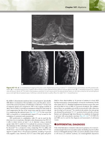
B C
Figure 107–15 A. T2-weighted axial imaging of thoracic spine. Myelomatous lesion involves T11 vertebral body and extends into left posterior ele-
ments. B. T1-weighted sequencing. Sagittal view of thoracic spine demonstrating diffuse marrow replacement. C. T2-weighted sequencing. Sagittal
view of thoracic spine. At T10, there is extraosseous extension of soft tissue within the paravertebral space on the right as well as the ventral epidural
space and right lateral epidural space. Soft tissue extends laterally to partly efface the right T10–11 neural foramen.
the ability to discriminate myeloma from normal marrow. Specifically, failed to show abnormalities in 30 percent of patients in whom MRI
MRI allows visualization of the medullary cavity and thus direct assess- had demonstrated an abnormal pattern of marrow involvement. On the
391
ment of the extent of myeloma cell infiltration of the bone. In the event other hand, PET-CT identified myelomatous lesions in areas that were
of suspected spinal cord compression, MRI is the imaging modality of out of the field of view of MRI in 35 percent of patients. The combina-
choice for its ability to provide an assessment of the level and extent of tion of these two modalities proved the most powerful with a detection
395
cord compression, the size of the tumor mass, and the extent to which rate of as high as 92 percent. In a multivariate analysis, the same group
it is compressing the epidural space (Fig. 107–15). In the event that also showed that persistent PET-CT positivity before and after primary
392
MRI is unavailable or contraindicated, urgent CT may be used for the therapy and subsequent high-dose therapy, is a predictor of prognosis
396
evaluation of a potential cord compression. in patients with symptomatic myeloma (Fig. 107–16). CT, PET-CT,
PET, particularly in combination with CT, can be used for the and MRI can be used for the diagnosis and assessment of soft-tissue
detection of active myeloma. Multiple studies demonstrate that PET-CT masses.
is able to detect lesions at least 1 cm in diameter using a standard
uptake value (SUV) cutoff of 2.5 to indicate the presence of disease. DIFFERENTIAL DIAGNOSIS
393
The limitation of this technology is that subcentimeter lesions may not
be detected. In a prospective study comparing PET-CT, MRI, and If the initial laboratory evaluation indicates the presence of a monoclo-
394
whole-body x-rays in newly diagnosed myeloma patients, PET-CT was nal immunoglobulin in serum and/or urine, the finding requires further
superior to plain films in 46 percent of patients, including 19 percent studies to distinguish among (1) MG; (2) solitary plasmacytoma of bone
with negative x-rays. However, PET-CT scans of the spine and pelvis or soft tissue; (3) indolent myeloma; (4) immunoglobulin deposition
Kaushansky_chapter 107_p1733-1772.indd 1749 9/21/15 12:35 PM