
Page 1944 - Williams Hematology ( PDFDrive )
P. 1944
1918 Part XII: Hemostasis and Thrombosis Chapter 113: Molecular Biology and Biochemistry of the Coagulation Factors 1919
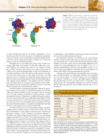
Arg 320 -IIe 321 Figure 113–4. Prothrombin to thrombin conversion.
A molecular model of prothrombin comprising the
Catalytic B chain γ-carboxy glutamic acid (Gla; GLA) domain, both kringle
domain domains, and the catalytic domain is shown (PDB struc-
A chain tures 2PF2, 1HAG, 1A0H, 1HAI). Gla domain-bound cal-
Arg 271 -Thr 272
cium ions are indicated in black. Cleavage by the factor
Va-Xa complex at Arg271 and Arg320 releases thrombin
(with the A chain and catalytically active B chain) from
the rest of the molecule (fragment 1.2).
Kringle 2
Thrombin
Kringle 1 Xa/Va
GLA
Prothrombin Fragment 1.2
at Arg155, thereby removing the Gla domain containing F1. The so in inflammatory and proliferative responses associated with vascular
formed meizo-des-F1 can be converted to thrombin by prothrombi- injury, such as in atherosclerosis and cancer. 26
nase, but at a slower rate as it is incapable of membrane binding. Assess- The physiologic inhibitors of thrombin are the serine protease
ment of F1.2 levels reflects prothrombin activation and is commonly inhibitors (serpins) antithrombin, heparin cofactor II, protein C inhib-
used as a marker for thrombin generation. itor, and protease nexin 1, with antithrombin being the primary plasma
Thrombin (IIα) is a two-chain serine protease (Mr ≈37,000) com- inhibitor. For all four serpins, the rate of thrombin inhibition can be
prising a light chain of 49 residues (A chain; Mr ≈6000) that is cova- accelerated by glycosaminoglycans, such as heparin (Table 113–4),
lently linked to the catalytic heavy chain of 259 residues (B chain; through mutual binding to the serpin and thrombin (see Fig. 113–2),
Mr ≈31,000). Thrombin’s main function is to induce the formation of which ensures rapid inhibition of thrombin at the intact endothelial cell
a fibrin clot by removing fibrinopeptides A and B from fibrinogen to surface where heparin-like glycosaminoglycans are found.
form fibrin monomers, which then spontaneously polymerize. In addi- Heparin and heparin-derivatives are clinically used as anticoagu-
tion, thrombin is able to cleave a wide variety of substrates with high lants to inhibit thrombin via antithrombin. Hirudin, which originates
specificity, which is mediated via its negatively charged, deep active site from the salivary glands of medicinal leeches, and its recombinant
cleft and via the anion binding exosites I and II that specifically interact and synthetic derivatives are potent and highly specific inhibitors
with cofactors and/or substrates. The dynamic structural conforma- that directly target the active site and exosite I of thrombin. The
27
19
tion of thrombin allows for binding to diverse ligands, and the subse- target-specific oral anticoagulant dabigatran also inhibits thrombin
quent ligand-induced conformational stabilization, known as thrombin directly with high specificity and reversibly binds the active site of
allostery, regulates and controls thrombin activity. 20,21 thrombin. 27,28
Thrombin initiates important procoagulant pathways by proteo-
lytic activation of the cofactors V and VIII and zymogen factor XI that
collectively amplify thrombin and fibrin formation, and by activating TABLE 113–4. Antithrombin Inhibition of Coagulation
factor XIII that crosslinks and stabilizes the fibrin polymers. Another Proteases
procoagulant function of thrombin is to inhibit fibrinolysis by proteo- Second-Order Association Rate Constants (M s )
–1 –1
lytic activation of the thrombin-activatable fibrinolysis inhibitor (TAFI), Protease – Heparin + H5 + UFH
a reaction enhanced by the endothelial-bound cofactor thrombomod-
ulin. Thrombin also has an anticoagulant function and upon binding to Thrombin 7.7 × 10 3 1.5 × 10 4 4.7 × 10 7
the cofactor thrombomodulin, it is capable of proteolytically activating Factor Xa 2.6 × 10 3 7.6 × 10 5 6.6 × 10 6
protein C, which inactivates the cofactors Va and VIIIa. Factor IXa 58 3.1 × 10 4 6.2 × 10 6
Thrombin activates the seven-transmembrane domain, G-protein–
coupled protease-activated receptors (PARs) PAR1, PAR3, and PAR4 TF-Factor VIIa 33 4.9 × 10 3 1.5 × 10 4
that are expressed on a wide range of cell types in the vasculature by Factor Xia 3.6 × 10 2 1.1 × 10 3 1.8 × 10 5
proteolytic cleavage of their N-terminal extracellular domains. 22–25 Factor XIIa 39 1.9 × 10 3 6.6 × 10 4
Thrombin is one of the strongest platelet activators in vivo and activates
platelet-expressed PAR1 and PAR4. The platelet glycoprotein (GP) Ib APC 0.08 1.9 2.1
25
(GPIb) serves as a cofactor for thrombin in PAR1 cleavage (Chap. 112). APC, activated protein C; TF, tissue factor.
Thrombin-mediated activation of endothelial-PAR1 triggers release of
von Willebrand factor (VWF) and P-selectin, which promote rolling The association rate constants characterizing the antithrombin inhi-
and adhesion of platelets and leukocytes. In addition, this stimulates the bition of coagulation proteases in the absence of heparin or accel-
erated by H5, the synthetic pentasaccharide fondaparinux, or UFH,
endothelial production of platelet-activating factor, a potent platelet and unfractionated heparin, which comprises long heparin molecules.
leukocyte activator, as well as the production of chemokines, cyclooxy- In vivo, natural glycosaminoglycan molecules on endothelium and
genase (COX)-2, and prostaglandins. Thrombin-mediated PAR activa- other cells accelerate the rate of inhibition. (See ST Olson, R Swanson,
25
tion is not only critical for coagulation, but also plays an important role E Raub-Segall, et al. )
321
Kaushansky_chapter 113_p1915-1948.indd 1919 9/21/15 2:39 PM