
Page 1948 - Williams Hematology ( PDFDrive )
P. 1948
1922 Part XII: Hemostasis and Thrombosis Chapter 113: Molecular Biology and Biochemistry of the Coagulation Factors 1923
is uncertainty about whether common gene variations influence the 12 3 456 7 8 9
level of factor X in plasma. 85 Gene 11 kb
PROTEIN C
Protein C, which plays a central role in the anticoagulant pathway,
was discovered in 1960, and being the third protein peak (“peak C”) mRNA 1.8 kb
observed in a vitamin K–dependent plasma protein purification, it was
named protein C. 86,87 Protein C is synthesized in the liver and circulates
in plasma as a two-chain zymogen of 417 amino acids (Mr ≈62,000) at a
concentration of 65 nM with a half-life of 6 to 8 hours (see Table 113–1). Exon 2 3 4 5 6 7 8 9
Protein Pro GLA EGF 1EGF 2 AP Catalytic domain
Protein Structure
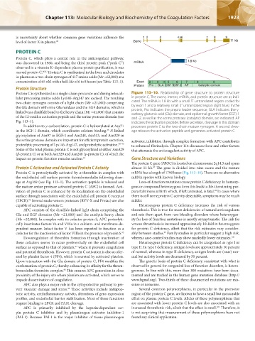
Protein C is synthesized as a single-chain precursor and during intracel- Figure 113–10. Relationship of gene structure to protein structure
lular processing amino acids Lys146-Arg147 are excised. The resulting in protein C. The exons, introns, mRNA, and protein structure are as indi-
two-chain zymogen consists of a light chain (Mr ≈21,000) comprising cated. The mRNA is 1.8 kb with a small 5′ untranslated region coded for
the Gla domain with nine Gla residues and the EGF domains, which is by exon 1 and a relatively small 3′ untranslated region (light blue). In the
linked via a disulfide bond to the heavy chain (Mr ≈41,000) that consists protein, Pro indicates the prepro leader sequence, GLA indicates the γ-
carboxy glutamic acid (Gla) domain, and epidermal growth factor (EGF)-1
of the 12-residue activation peptide and the serine protease domain (see and -2, as well as the serine protease (catalytic) domain, are indicated. AP
Fig. 113–1). indicates the activation peptide. Before secretion, cleavage in this domain
In addition to γ-carboxylation, protein C is hydroxylated at Asp71 processes protein C to the two-chain mature zymogen. A second cleav-
in the EGF-1 domain, which coordinates calcium binding. N-linked age releases the activation peptide and generates activated protein C.
35
glycosylation of Asn97 in EGF-1 and Asn248, Asn313, and Asn329 in
the serine protease domain are important for efficient protein secretion, activator, inhibition through complex formation with APC contributes
proteolytic processing of Lys146-Arg147, and proteolytic activation. 88–90 to enhanced fibrinolysis. Chapter 114 discusses these and other factors
Some of the total plasma protein C is not glycosylated at either Asn329 that attenuate the anticoagulant activity of APC.
(β-protein C) or at both Asn329 and Asn248 (γ-protein C), of which the
impact on protein function remains unclear. 91 Gene Structure and Variations
The protein C gene (PROC) is located on chromosome 2q14.3 and spans
Protein C Activation and Activated Protein C Activity almost 11 kb. The gene is divided into nine exons and the mature
97
Protein C is proteolytically activated by α-thrombin in complex with mRNA has a length of 1790 bases (Fig. 113–10). There are no alternative
the endothelial cell surface protein thrombomodulin following cleav- mRNA species with known biology.
age at Arg169 (see Fig. 113–1). The activation peptide is released and Loss-of-function mutations cause protein C deficiency. In homozy-
the mature serine protease activated protein C (APC) is formed. Acti- gous or compound heterozygous form this leads to life-threatening pur-
vation of protein C is enhanced by its localization on the endothelial pura fulminans at birth which, if left untreated, is fatal. In cases where
98
surface through association with the endothelial cell protein C receptor there is still some protein C activity detectable, symptoms may be much
(EPCR). Several snake venom proteases (RVV-X and Protac) are also milder.
92
capable of activating protein C. Heterozygous protein C deficiency increases the risk of venous
APC consists of the disulfide-linked light chain comprising the thrombosis. This is true for most deficiencies of natural anticoagulants
Gla and EGF domains (Mr ≈21,000) and the catalytic heavy chain and sets them apart from rare bleeding disorders where heterozygos-
(Mr ≈32,000). In complex with its cofactor protein S, APC proteolyti- ity for loss of function mutations is mostly asymptomatic. The risk for
cally inactivates factors Va and VIIIa in a calcium- and membrane-de- venous thrombosis is increased approximately 10-fold in heterozygotes
pendent manner. Intact factor V has been reported to function as a for protein C deficiency, albeit that the risk estimates vary consider-
cofactor for the inactivation of factor VIIIa in the presence of protein S. 93 ably between studies. Family studies in particular suggest a high risk,
99
Downregulation of thrombin formation through inactivation of whereas case-control studies may show markedly lower estimates. 100
these cofactors seems to occur preferentially on the endothelial cell Heterozygous protein C deficiency can be categorized as type I or
surface as opposed to that of platelets, where it prevents coagulation type II. In type I deficiency, antigen levels are approximately 50 percent
94
and potential thrombosis. However, protein C activation is also acceler- of normal, whereas in type II deficiency, antigen levels are (near) nor-
ated by platelet factor 4 (PF4), which is secreted by activated platelets. mal but activity levels are decreased by 50 percent.
Upon interaction with the Gla domain of protein C, PF4 modifies the The genetic basis of protein C deficiency, consistent with what is
conformation of protein C, thereby enhancing its affinity for the throm- observed in general for congenital loss of function disorders, is hetero-
bomodulin-thrombin complex. This ensures APC generation in close geneous. In line with this, more than 300 mutations have been docu-
95
proximity of the injury site where platelets are activated, which serves to mented and are tracked in the human gene mutation database (http://
impede dissemination of coagulation. www.hgmd.org). Two-thirds of these documented mutations are mis-
APC also plays a major role in the cytoprotective pathway to pre- sense or nonsense.
vent vascular damage and stress. These activities include antiapop- Several common polymorphisms, in particular in the promotor
96
totic activity, antiinflammatory activity, alterations of gene-expression region of the protein C gene, are known to have a small but measurable
profiles, and endothelial barrier stabilization. Most of these functions effect on plasma protein C levels. Alleles of these polymorphisms that
require binding to EPCR and PAR1 cleavage. are associated with lower protein C levels are also associated with an
APC is primarily inhibited by the heparin-dependent ser- increased thrombotic risk, albeit that the effect is small. Therefore, it
101
pin protein C inhibitor and by plasminogen activator inhibitor-1 is not surprising that measurement of these polymorphisms have not
(PAI-1). Because PAI-1 is the major inhibitor of tissue plasminogen found any clinical application.
Kaushansky_chapter 113_p1915-1948.indd 1923 9/21/15 2:39 PM