
Page 1947 - Williams Hematology ( PDFDrive )
P. 1947
1922 Part XII: Hemostasis and Thrombosis Chapter 113: Molecular Biology and Biochemistry of the Coagulation Factors 1923
A defect or deficiency in factor IX leads to hemophilia B. Chap- of factor Xa at adjacent C-terminal Arg or Lys residues also results in
ter 123 discusses the prevalence, clinical characteristics, and molecular the generation of factor Xaβ and factor Xaβ derivatives. 73,74 While the
genetics of hemophilia B in detail. coagulation activity is eliminated in the factor Xaβ derivatives, they are
Conversely, increased levels of factor IX are a strong risk fac- capable of interacting with the zymogen plasminogen and enhance its
tor for venous thrombosis. This is in agreement with a rare gain of tissue plasminogen activator-mediated conversion to plasmin, thereby
58
function mutation (Arg335Leu; factor IX Padua), which renders the promoting fibrinolysis. 75
protein hyperfunctional and is associated with familial early-onset A primary plasma inhibitor of factor Xa is the serpin anti-
thrombophilia. 59 thrombin, and this inhibition is enhanced by heparin (see Table 113–4),
which induces a conformational change in antithrombin that is required
for simultaneous active site and exosite interactions with factor Xa.
76
FACTOR X Another potent factor Xa inhibitor is TFPI, which inhibits both the
Factor X was originally reported in the late 1950s as the “Stuart-Prower ternary tissue factor–factor VIIa–factor Xa complex as well as free fac-
factor,” named after the first two identified factor X–deficient patients. 60–62 tor Xa, for which protein S functions as a cofactor. 77,78 Free factor Xa is
Factor X is primarily synthesized in the liver and circulates in plasma as also inhibited by the protein Z/protein Z–dependent protease inhibitor
a two-chain zymogen of 445 amino acids (Mr ≈59,000) at a concentra- (ZPI) complex on membranes. 79
tion of 170 nM with a half-life of 34 to 40 hours (see Table 113–1). Low-molecular-weight heparin and synthetic derivatives (e.g.
fondaparinux) are clinically used as anticoagulants to enhance factor
Protein Structure Xa inhibition by antithrombin specifically. The target-specific oral anti-
Factor X is synthesized as a single-chain precursor and during intracel- coagulants rivaroxaban, apixaban, edoxaban, and analogues directly
lular processing, the three-amino acid peptide Arg140-Lys141-Arg142 inhibit both free factor Xa and prothrombinase complex-assembled
is excised. The resulting two-chain zymogen consists of a light chain factor Xa with high specificity through a high-affinity, reversible inter-
(Mr ≈16,000), comprising the Gla domain with 11 Gla residues and the action with the factor Xa active site. 80–83
EGF domains, which is linked via a disulfide bond to the heavy chain
(Mr ≈42,000) that consists of a 52-residue activation peptide and the Gene Structure and Variations
serine protease domain (see Fig. 113–1). The gene encoding factor X (F10) is located on chromosome 13q34 and
84
Hydroxylation of Asp63 mediates calcium binding to the EGF spans almost 27 kb. The 8 exons in the factor X gene give rise to a
1 domain and orients the Gla domain, which is essential for factor X mature mRNA of 1560 bases (Fig. 113–9). There are no common alter-
clotting activity. N-linked glycosylation of the activation peptide resi- native splice variants with known biology.
35
dues Asn181 and Asn191 has been implicated in prolonging the factor Loss of function mutations in the factor X gene lead to a rare bleed-
X half-life. Further posttranslational modification of factor X consists ing disorder with a recessive mode of inheritance. Factor X deficiency
63
of O-linked glycosylation at Thr159 and Thr171 in the activation pep- occurs in approximately one in every 1,000,000 newborns. Most cases of
tide and Thr443 in the serine protease domain. There is some evidence documented factor X deficiency experience serious bleeding problems.
that glycosylation of the human factor X activation peptide may also In fact, factor X deficiency may be the most severe of the rare congenital
30
contribute to substrate recognition by the intrinsic or extrinsic factor bleeding disorders. Well over 100 mutations have been documented in
X-activating complex. 64,65 cases with factor X deficiency (http://www.hgmd.org). The majority of
these mutations are missense and nonsense mutations.
Factor X Activation and Factor Xa Activity Gain-of-function mutations in factor X could potentially increase
Factor X is proteolytically activated by either the factor VIIIa–factor IXa thrombotic risk, but such mutations have not been documented. There
(“intrinsic tenase”) or the tissue factor–factor VIIa (“extrinsic tenase”)
enzyme complexes following cleavage at Arg194 in the heavy chain (see
Fig. 113–1). This results in the release of the activation peptide and gen- 1 2 34 5 6 7 8
eration of factor Xa, also known as factor Xaα. A snake venom protease Gene 25 kb
from Russell’s viper venom (RVV-X) is capable of generating factor Xa
in a similar manner.
Factor Xa consists of the Gla and EGF domains-comprising light
chain (Mr ≈16,000) and the catalytic heavy chain (Mr ≈29,000) that are mRNA 1.5 kb
covalently linked via a disulfide bond. Factor Xa reversibly associates
with its cofactor factor Va on an anionic membrane surface in the pres-
ence of calcium ions to form prothrombinase, the physiologic activator
of prothrombin. Factor Xa is also involved in the proteolytic activation Exon
6
5
34
8
of factors V, VII, and VIII. 66–68 Protein 1 Pro 2 GLA EGF 1EGF 2 AP 7 Catalytic domain
Similar to thrombin, factor Xa plays a role in other biologic and
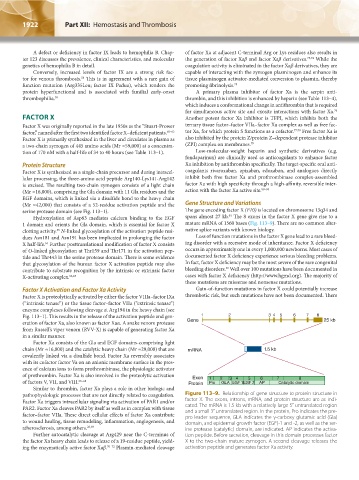
pathophysiologic processes that are not directly related to coagulation. Figure 113–9. Relationship of gene structure to protein structure in
Factor Xa triggers intracellular signaling via activation of PAR1 and/or factor X. The exons, introns, mRNA, and protein structure are as indi-
PAR2. Factor Xa cleaves PAR2 by itself as well as in complex with tissue cated. The mRNA is 1.5 kb with a relatively large 5′ untranslated region
factor–factor VIIa. These direct cellular effects of factor Xa contribute and a small 3′ untranslated region. In the protein, Pro indicates the pre-
pro leader sequence, GLA indicates the γ-carboxy glutamic acid (Gla)
to wound healing, tissue remodeling, inflammation, angiogenesis, and domain, and epidermal growth factor (EGF)-1 and -2, as well as the ser-
atherosclerosis, among others. 26,69 ine protease (catalytic) domain, are indicated. AP indicates the activa-
Further autocatalytic cleavage at Arg429 near the C-terminus of tion peptide. Before secretion, cleavage in this domain processes factor
the factor Xa heavy chain leads to release of a 19-residue peptide, yield- X to the two-chain mature zymogen. A second cleavage releases the
ing the enzymatically active factor Xaβ. 70–72 Plasmin-mediated cleavage activation peptide and generates factor Xa activity.
Kaushansky_chapter 113_p1915-1948.indd 1922 9/21/15 2:39 PM