
Page 1949 - Williams Hematology ( PDFDrive )
P. 1949
1924 Part XII: Hemostasis and Thrombosis Chapter 113: Molecular Biology and Biochemistry of the Coagulation Factors 1925
THE PROCOAGULANT COFACTORS Protein Structure
V AND VIII Factor V has an A1-A2-B-A3-C1-C2 domain structure (Fig. 113–11).
The three A-type domains share significant homology with those
of ancestral ceruloplasmin as well as with the factor VIII A domains
Factors V and VIII both function as cofactors in coagulation and dra-
matically enhance the catalytic rate of their macromolecular enzyme (approximately 50 percent sequence identity). The two C-type domains
complexes, resulting in the generation of thrombin and factor Xa, respec- belong to the family of discoidin domains, which are generally involved
tively. Apart from their functional equivalence, they also share similar in cell adhesion, and share approximately 55 percent sequence identity
gene structures, amino acid sequences, and protein domain structures, with the factor VIII C domains. The C domains mediate binding to the
which is not surprising considering that factors V and VIII are assumed anionic phospholipid surface, thereby localizing factor V to the site of
115–118
to descend from the common ancestral A1-A2-A3 domain-containing injury and facilitating interaction with factor Xa and prothrombin.
copper-binding plasma protein ceruloplasmin through a gene duplica- In contrast, the large central B domain of factor V shows weak homol-
tion event. After acquiring C-type domains as well as the central B ogy to the factor VIII B domain or to any other known protein domain.
102
domain, a second gene duplication ultimately separated the ancestral However, this domain comprises so-called basic and acidic regions that
genes of factors V and VIII. are highly conserved throughout evolution and serve to negatively regu-
119,120
Factors V and VIII undergo similar mechanisms of intracellular late factor V function and prevent activity of the procofactor.
processing in the endoplasmic reticulum (ER) and Golgi apparatus. Factor V undergoes extensive posttranslational modifications,
35,121
Trafficking through this early secretory pathway involves interaction of including sulfation, phosphorylation, and N-linked glycosylation.
factors V and VIII with a receptor complex that consists of the man- Sulfation at sites in the A2 and B domain are involved in the thrombin-
122
nose-binding lectin-1 gene product LMAN1 (also called ER-Golgi mediated proteolytic activation of factor V. Phosphorylation at Ser692
intermediate compartment (ERGIC)-53) and multiple coagulation defi- in the A2 domain enhances the APC-dependent inactivation of the
123
ciency protein 2 (MCFD2). Defects or deficiencies in one of the two cofactor Va. N-linked glycosylation occurs throughout the whole pro-
103
subunits of the receptor complex can result in a combined deficiency of tein; however, the majority of carbohydrates are linked to Asn residues
factors V and VIII (Chap. 124). within the B domain and play a role in the LMAN1-MCDF2 receptor
complex-mediated trafficking of factor V from the ER to the Golgi in
the early secretory pathway. Partial glycosylation at Asn2181 in the
103
FACTOR V C2 domain of factor V results in a lower binding affinity for negatively
In 1943, Norwegian physician Paul Owren discovered the fifth coag- charged membranes of the glycosylated form, thereby reducing the fac-
ulation factor thus far known and named it factor V. 104–106 Factor V is tor V procoagulant activity, particularly at low phospholipid concen-
synthesized in the liver and circulates in plasma as a large single-chain trations. 124,125 Furthermore, factor V comprises several disulfide bonds
procofactor of 2196 amino acids (Mr ≈330,000) at a concentration of 20 that are important for the three-dimensional structure of the A and C
nM with a half-life of 12 to 36 hours (see Table 113–1). domains. 121
Approximately 20 percent of the total factor V in blood is stored
in the α-granules of platelets. Although it was originally thought that Factor V Procofactor Activation and Factor Va Cofactor
megakaryocytes synthesize factor V, studies in humans indicate that Function
platelet factor V originates from plasma through endocytic uptake. 107–109 Sequential proteolytic cleavage of the procofactor factor V at Arg709,
Platelet factor V is modified intracellularly such that it is functionally Arg1018, and Arg1545 in the B domain results in release of the inhib-
unique compared to its plasma-derived counterpart. It is partially acti- itory constraints exerted by the B domain and in the generation of the
vated, more resistant to inactivation by APC, and has several different heterodimeric cofactor Va (see Fig. 113–11). Maximal cofactor activ-
126
posttranslational modifications. 110 ity correlates with cleavage at Arg1545, which is consistent with the
Platelet factor V is associated with the large multimeric protein observation that a snake venom protease from RVV-V, which cleaves
multimerin. Multimerin has a massive repeating structure, with some only at Arg1545, results in full activation. Thrombin has generally been
111
of the multimers having molecular weights of several million daltons. recognized to be the principal activator of factor V. However, recent
Although the function of this platelet factor V-specific multimeric findings suggest that in the initiation phase of coagulation factor V is
chaperon protein is similar to that of VWF, the multimeric chaperon primarily activated by factor Xa. Factor Xa initially cleaves factor V at
127
protein of factor VIII in plasma, multimerin and VWF share no struc- Arg1018, followed by proteolysis at Arg709 and Arg1545. 128
tural homology. Factor Va is composed of a heavy chain (Mr ≈105,000) compris-
Following platelet activation, platelet factor V becomes available ing the A1-A2 domains and the A3-C1-C2 light chain (Mr ≈74,000),
at the site of injury and can reach local concentrations that exceed the which are noncovalently associated via calcium ions. Factor Va is a
factor V plasma concentration by more than 100-fold. Interestingly, nonenzymatic cofactor within the prothrombinase complex that greatly
112
the origin of factor V in mouse platelets differs from humans in that it accelerates the ability of factor Xa to rapidly convert prothrombin to
is synthesized in megakaryocytes and stored into the α-granules before thrombin. APC catalyzes the inactivation of factor Va by cleavage at
129
platelets are released from the marrow. 113,114 the main sites Arg306 and Arg506, upon which the cleaved A2 fragment
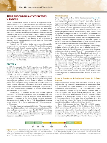
A1 A2 B A3 C1 C2
306 506 709 1018 1545
Figure 113–11. The domain structure of factor V. Schematic A1-A2-B-A3-C1-C2 domain representation of factor V. Thrombin cleavage sites (Arg709,
Arg1018, Arg1545) are indicated by green arrows, and activated protein C (APC) cleavage sites (Arg306, Arg506) by red arrows. The blue and red boxes
in the B domain represent the basic and acidic region, respectively, that are highly conserved throughout evolution and serve to negatively regulate
factor V function and prevent activity of the procofactor V.
Kaushansky_chapter 113_p1915-1948.indd 1924 9/21/15 2:39 PM