
Page 1945 - Williams Hematology ( PDFDrive )
P. 1945
1920 Part XII: Hemostasis and Thrombosis Chapter 113: Molecular Biology and Biochemistry of the Coagulation Factors 1921
12 34 56 78 9101112 1314 and N-linked (Asn154 in the connecting region, Asn322 in the serine
Gene 20 kb protease domain) glycosylation.
Factor VII Activation and Factor VIIa Activity
Factor VII is proteolytically activated once it has formed a high-affin-
ity complex with its cofactor tissue factor. A number of coagulation
mRNA 2 kb
proteases including thrombin and factors IXa and XIIa are capable of
cleaving factor VII at Arg152 to generate factor VIIa (see Fig. 113–1),
with factor Xa being considered the most potent and physiologically
36
relevant activator of factor VII. Autoactivation can also occur, which
Exon 1 2 34 5 6 7 8 9 10 11 12 1314 is initiated by minute amounts (approximately 0.1 nM) of preexisting
Protein Pro GLA Kringle 1 Kringle 2 LC Catalytic domain
factor VIIa. 37
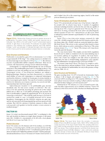
Figure 113–5. Relationship of gene structure to protein structure in Factor VIIa is a two-chain serine protease composed of a light
prothrombin. The exons, introns, mRNA, and protein structure are as chain (Mr ≈20,000) comprising the Gla and EGF domains and the cata-
indicated. The mRNA is 2 kb with small 5′ and 3′ untranslated regions lytic heavy chain (Mr ≈30,000), which are covalently linked via a disul-
(shown in light blue). In the protein, Pro indicates the prepro leader fide bond. Factor VIIa activity is only expressed when bound to tissue
sequence, GLA indicates the γ-carboxy glutamic acid (Gla) domain, factor, which induces an active conformation of the factor VIIa serine
Kringles 1 and 2 are indicated, LC indicates the light chain (also known protease domain (Fig. 113–6). Factor VIIa interacts with tissue factor
11
as the A chain), and the serine protease (catalytic) domain is indicated.
via its Gla and EGF domains.
The tissue factor–factor VIIa complex activates both coagulation
factors IX and X, which is considered to be the main initiating step of
Gene Structure and Variations the extrinsic pathway of coagulation. In addition, the tissue factor–
Prothrombin is encoded by a gene (F2) on chromosome 11p11.2 that factor VIIa (–factor Xa) complex is not only critical to processes in
is approximately 20 kb long. The coding sequence is divided over coagulation, but also to wound healing, angiogenesis, tissue remodel-
29
14 exons that range in size from 25 to 315 bp (Fig. 113–5). The reference ing, and inflammation through proteolytic activation of PAR2. 38–40
sequence of prothrombin mRNA comprises 2018 bases. There are no The ternary tissue factor–factor VIIa–factor Xa complex is inhib-
common, well characterized, splicing variants with known biology. ited by the tissue factor pathway inhibitor (TFPI). Tissue factor–factor
Homozygosity or compound heterozygosity for loss of function VIIa is also inhibited by antithrombin, but only in the presence of hep-
mutations in the prothrombin gene leads to a bleeding tendency. This arin (see Table 113–4).
condition is quite rare with perhaps one case per 2,000,000 newborns.
30
Heterozygous carriers of loss-of-function mutations are without a Gene Structure and Variations
bleeding phenotype. Mutations have been characterized in a relatively The gene encoding factor VII (F7) is located on chromosome 13q34,
small number of cases with homozygous or compound heterozygous is almost 15 kb in length, and comprises 9 exons (Fig. 113–7). The
prothrombin deficiency (consult the human gene mutation database at canonical mRNA encoding factor VII comprises 3000 bases. Alterna-
41
http://www.hgmd.org for details). The majority of mutations underlying tively spliced transcript variants encoding multiple isoforms have been
prothrombin deficiency are missense mutations, but several small dele- observed, but their biology is not well characterized. 42
tions/insertions have also been reported. Inherited factor VII deficiency is a rare autosomal recessive dis-
Gain-of-function mutations in the prothrombin gene increase order that affects approximately one in 500,000 newborns. Factor VII
30
thrombotic risk. The best known variation is G20210A. This vari- deficiency is the most common of the inherited rare bleeding disorders,
31
ation of the last nucleotide preceding the poly(A)-tail of the mature
mRNA has an effect on 3′-end mRNA processing and increases the
level of prothrombin in plasma by approximately 10 to 20 percent in Catalytic
heterozygous individuals. This relatively small increase in the level of
32
prothrombin results in a two- to threefold enhanced risk for venous
thrombosis. Homozygotes for the G20210A variation are quite rare,
and the risk associated with homozygosity has not been measured with
certainty. The G20210A variation is relatively common in whites, with EGF 2
a strong south-north gradient in that the variation is most common in
southern Europe. 33 EGF 1
FACTOR VII GLA
Factor VII, which was discovered around 1950, is synthesized in the
34
liver and circulates in plasma as a single-chain zymogen of 406 amino
acids (Mr ≈50,000) at a concentration of 10 nM with a short plasma Factor Vlla Tissue factor
half-life of 3 to 6 hours (see Table 113–1).
Figure 113–6. The tissue factor–factor VIIa complex. A molecular
Protein Structure model of factor VIIa (PDB structures 1QHK, 1WHF, 1RFN, 1DAN) and the
Factor VII consists of a Gla domain with 10 Gla residues, two EGF-like crystal structures of the tissue factor–factor VIIa complex (PDB struc-
ture 1DAN) and the extracellular domain of tissue factor (PDB structure
domains, a connecting region, and the serine protease domain (see Fig. 2HFT) are shown. The γ-carboxy glutamic acid (Gla; GLA) domain, epi-
113–1). Calcium coordination in EGF-1 is mediated by partial hydroxy- dermal growth factor (EGF)-like domains 1 and 2, and serine protease
lation of Asn63 and O-linked fucosylation of Ser60. Further posttrans- (catalytic) domain of factor VIIa are indicated. Binding to tissue factor
35
lational modifications of factor VII consist of O-linked (Ser52 in EGF-1) alters the overall structure of factor VIIa.
Kaushansky_chapter 113_p1915-1948.indd 1920 9/21/15 2:39 PM