
Page 1957 - Williams Hematology ( PDFDrive )
P. 1957
1932 Part XII: Hemostasis and Thrombosis Chapter 113: Molecular Biology and Biochemistry of the Coagulation Factors 1933
1 Endothelial Protein C Receptor Function
Gene 3.7 kb EPCR enhances the activation of membrane-bound protein C by the
thrombomodulin–thrombin complex, thereby enhancing the APC-
92
mediated anticoagulant pathway.
APC bound to membrane-associated or soluble EPCR is disabled
mRNA 3.7 kb in its anticoagulant capacity. Instead, EPCR-bound APC activates PAR1
in an alternative manner by noncanonical cleavage at a Arg46, result-
281
ing in an increased barrier function of endothelial cells mediated via
the β-Arrestin/PI3K (phosphatidylinositide 3′-kinase)/AKT/Rac1 path-
way. This is in contrast to the barrier-disruptive Arg41 cleavage of PAR1
Exon 1 by thrombin that activates the G-protein/ERK (extracellular regulated
Protein Pro Lectin-like domain E1 E2 E3 E4 E5 E6 S/T MCyt
kinase) 1.2/RhoA pathway. 281
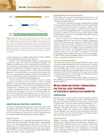
Figure 113–19. Relationship of gene structure to protein structure EPCR is essential at the maternal–embryonic interface on tro-
in thrombomodulin. The thrombomodulin gene has no introns. The phoblast giant cells where it prevents fibrin formation. Consequently,
exon, mRNA, and protein structure are as indicated. The mRNA is 3.7 kb, complete EPCR deficiency leads to embryonic lethality. EPCR-deficient
with a small 5′ untranslated region and a large 3′ untranslated region embryos rescued by the presence of EPCR in the trophoblast are viable
(light blue). In the protein, Pro indicates the prepro leader sequence, the and thrive, which seems to indicate that EPCR is not essential to blood
lectin-like domain is indicated, E indicates the epidermal growth factor circulation, at least in mice. Additional ligands for EPCR have been
282
(EGF)-like domains, S/T indicates the serine- and threonine-rich region,
M indicates the transmembrane region, and Cyt indicates the cytoplas- discovered such as factor VIIa, Plasmodium falciparum erythrocyte
283
mic domain. membrane protein, and the V(γ)4V(δ)5 T-cell receptor. These addi-
tional ligands indicate potential involvement of EPCR in the therapeu-
tic effect of factor VIIa in hemophilia patients, and roles for EPCR in
mutations, however, do not explain a large proportion of the heritability malaria, cytomegalovirus infection, and cancer.
of venous thrombosis as they seem to be quite rare.
Recently a novel thrombomodulin mutation, p.Cys537Stop, was Gene Structure and Variations
described in a family with a history of posttraumatic bleeding. The The chromosomal location of the EPCR gene (PROCR) is 20q11.2 and it
277
endogenous thrombin potential was markedly reduced at low tissue fac- contains four exons and spans 6 kb. Exon 1 encodes for the 5′-untrans-
tor concentrations in heterozygous carriers. Plasma thrombomodulin lated region and the signal peptide; exons 2 and 3 encode for almost the
levels were elevated (433 to 845 ng/mL, normal range 2 to 8 ng/mL), entire extracellular region; and exon 4 encodes for the transmembrane
and the addition of exogenous protein C further decreased thrombin domain and cytoplasmic tail. One single mRNA encodes the centroso-
generation. It was surmised that as a consequence of the premature stop mal protein CCD41 and EPCR. Deletion of the signal sequence confers
codon, the truncated thrombomodulin is shed from the endothelial sur- the centrosomal location of CCD41, while the unprocessed protein is
face into the blood plasma, which would promote systemic protein C incorporated into cell membranes as EPCR.
activation, thereby explaining the bleeding phenotype. Variants of EPCR with reduced protein C affinity or increased cel-
Missense mutations in thrombomodulin were also reported lular shedding are reported to be associated with unprovoked venous
in patients with aHUS and this involvement in aHUS is probably thromboembolism. 284
related to the role of thrombomodulin in the complement system.
278
Thrombomodulin binds to C3b and factor H and negatively regulates THE FIBRIN NETWORK: FIBRIN(OGEN),
complement by accelerating factor I-mediated inactivation of C3b.
In addition, by promoting activation of TAFI, thrombomodulin also FACTOR XIII, AND THROMBIN-
accelerates the inactivation of C3a and C5a. Thrombomodulin vari- ACTIVATABLE FIBRINOLYSIS INHIBITOR
ants associated with aHUS had diminished capacity to inactivate C3b
and to activate TAFI and were thus less protected from activated com- FIBRINOGEN
plement, thereby providing an explanation for their involvement in Fibrinogen, when converted to fibrin, forms the structural meshwork
aHUS.
that consolidates an initial platelet plug into a solid hemostatic clot.
Fibrinogen is synthesized in the liver and circulates in a concentration
ENDOTHELIAL PROTEIN C RECEPTOR of approximately 7.4 μM. The plasma half-life of fibrinogen is 3 to 5
days, with only a small proportion of the catabolism caused by con-
The EPCR, a single-chain transmembrane receptor discovered in 1995 sumption. Fibrinogen is also found in the α-granules of platelets. It
285
by Fukodome and Esmon, binds both protein C and APC. EPCR was initially assumed that megakaryocytes synthesized fibrinogen.
279
increases the rate of activation of protein C and alters the function of However, although some γ-chain transcripts are present in marrow pre-
92
APC from anticoagulant to cytoprotective. EPCR is mainly expressed cursors, it appears that most of the fibrinogen found within platelets is
280
by endothelial cells but also by leukocytes and other cell types. taken up from the plasma by endocytosis. 286,287
Protein Structure Protein Structure
EPCR is homologous to CD1 and major histocompatibility class I pro- Chapter 135 provides a detailed description of the biochemistry of
teins and folds with a β-sheet platform supporting two α-helical regions fibrinogen and of fibrin formation and degradation. Fibrinogen is a
that form the potential binding pocket for protein C and APC. The dimeric GP (Mr ≈340,000) and each of the two subunits contains three
mature protein (Mr ≈49,000) consists of 223 amino acids and is glycosy- disulfide-linked polypeptide chains that are referred to as the Aα (Mr
lated through four N-linked glycosylation sites (Asn30, Asn47, Asn119, ≈66,500), Bβ (Mr ≈52,000), and γ (Mr ≈46,500) chains. A trinodular
Asn155). EPCR contains a 25-residue long C-terminal transmembrane model of fibrinogen structure has been established from the crystal
region with a short 3-residue cytoplasmic tail. structure of fibrinogen (Fig. 113–20). 288
Kaushansky_chapter 113_p1915-1948.indd 1932 9/21/15 2:40 PM