
Page 1956 - Williams Hematology ( PDFDrive )
P. 1956
1930 Part XII: Hemostasis and Thrombosis Chapter 113: Molecular Biology and Biochemistry of the Coagulation Factors 1931
1 2 3 4 5 6 Protein Structure
12 kb
Gene 12 kb Mature single-chain thrombomodulin comprises 557 residues (Mr
≈78,000) and is composed of a lectin-like domain, a hydrophobic
region, six EGF-like domains, a serine- and threonine-rich region, a
transmembrane domain, and a 23-residue cytoplasmic tail. The highly
2.3 kb
mRNA 2.3 kb charged lectin-like domains bear homology to the C-type lectins. Post-
translational modifications include five N-linked glycosylation sites that
are located in the lectin-like and EGF-4 and -5 domains. O-linked gly-
cosylation in the serine- and threonine-rich region (Ser474) supports
attachment of a glycosaminoglycan, a chondroitin sulfate moiety, which
Exon 1 2 3 4 5 6 forms a low-affinity binding site for thrombin.
Protein Pro Extracellular domain Tran Cyto
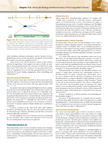
Figure 113–18. Relationship of gene structure to protein structure Thrombomodulin Cofactor Function
in tissue factor. The exons, introns, mRNA, and protein structure are Thrombomodulin interacts with thrombin through its EGF-5 and -6
as indicated. The mRNA is 2.3 kb with a 5′ untranslated region and a domains in a calcium-dependent manner. As a result, thrombin’s pro-
267
large 3′ untranslated region (light blue). Pro indicates the prepro leader coagulant exosite I is shielded, which causes thrombin’s specificity to
sequence, the extracellular domain is indicated, Tran indicates the trans-
membrane region, and Cyto indicates the cytoplasmic domain. switch to the anticoagulant substrate protein C, requiring EGF domains
4 to 6 of thrombomodulin, and to TAFI, requiring EGF-3 to -6. Throm-
268
bomodulin enhances the thrombin-dependent activation of protein C
more than 1000-fold.
redox (oxidation-reduction) environment and the exposure of phos- As a result of the relatively large endothelial surface area in cap-
phatidylserine. However, both monomeric and dimeric forms of tissue illary beds, the thrombomodulin-dependent activation of protein C
factor appear to possess procoagulant activity. 255 proceeds efficiently in the microcirculation, which serves a major role
Tissue factor is not only the primary initiator of the extrinsic in preventing thrombosis from occurring on intact endothelium. In
269
pathway of coagulation, it also supports activation of PAR2 on endo- larger vessels where the endothelial surface area-blood volume ration is
thelial cells and smooth muscle cells. Activation of PAR2 by the tissue low, the presence of EPCR aids in the interaction with and presentation
factor–factor VIIa(–factor Xa) complex is not necessarily directly rel- of protein C to the thrombomodulin-thrombin complex. 270
evant for coagulation, but it is currently speculated that this event is Thrombomodulin also enhances the thrombin-mediated con-
important for wound healing, angiogenesis, tissue remodeling, and version of single-chain urokinase-type plasminogen activator to
inflammation. 38–40 thrombin-cleaved two-chain urokinase-type plasminogen activa-
tor, which interferes with the generation of plasmin. Furthermore,
Gene Structure and Variations thrombomodulin is a negative regulator of PAR signaling, as throm-
The human tissue factor gene is located on chromosome 1p21-p22. bomodulin-bound thrombin is incapable of PAR activation. Based
271
The DNA sequence of the tissue factor gene has been determined and on this and because thrombomodulin is the cofactor responsible for
consists of six exons and five introns that span approximately 12 kb APC generation, thrombomodulin plays an important role, albeit
(Fig. 113–18). indirect, as an antiinflammatory protein. A direct contribution to sup-
The primary transcript encoding full-length tissue factor contains press inflammation has been attributed to the lectin-like domains and
six exons, but an alternatively spliced form of tissue factor (asTF) also hydrophobic region of thrombomodulin, independent of its anticoag-
exists in which exon 5 is spliced out. Because of a 3′ frameshift muta- ulant activity. 272
tion, the full-length tissue factor transmembrane and cytoplasmic tail Protein C inhibitor is an effective inhibitor of the thrombomod-
are replaced with a hydrophobic C-terminal domain, which renders the ulin-thrombin complex. 273
asTF soluble. asTF is expressed in lung, pancreas, placenta, heart, endo- Proteolysis of thrombomodulin by neutrophil-derived metallo-
thelium, and monocytes. 259–261 Although the level of asTF in human proteinases and possibly rhomboids results in the generation of soluble
plasma may be substantial and amounts to 10 to 30 percent of total tis- thrombomodulin. Normal plasma levels of soluble thrombomodulin
274
sue factor, it remains a matter of debate whether asTF contributes to are 3 to 50 ng/mL, but may increase as a result of vascular damage asso-
262
coagulation. ciated with infection, sepsis, or inflammation. 274
In theory, variations in the tissue factor gene could influence
thrombotic and bleeding risk. There are claims that polymorphisms in Gene Structure and Variations
the tissue factor gene influence thrombotic risk but these claims have The human thrombomodulin gene (THBD) is located on chromo-
not been sufficiently confirmed. 263 some 20p11.2, spans approximately 3.5 kb, and consists of a single
No relationship between loss-of-function mutations and bleeding exon (Fig. 113–19). Intronless genes are uncommon in eukaryotes and
has been described. This is perhaps not surprising in view of the fact include rhodopsin, angiogenin, mitochondrial genes, interferons α- and
that mice lacking tissue factor die early in gestation. β-adrenergic receptors. Intronless genes represent recent additions to
the genome, created mostly by retroposition of processed mRNAs with
retained functionality. Genetic variation in thrombomodulin has been
THROMBOMODULIN studied in conjunction with venous thrombosis, bleeding and atypical
Thrombomodulin, which was first identified by Esmon and colleagues hemolytic uremic syndrome (aHUS).
in the early 1980s, 264,265 is a predominantly endothelial transmembrane There are early reports that mutations in thrombomodulin are
protein and functions as an endothelial receptor for thrombin. In addi- present in patients with venous thrombosis, but it was difficult to
275
tion to endothelium, thrombomodulin has also been detected on a prove causality. More recent work that made use of thrombomodulin
number of other cell types, including megakaryocytes, monocytes, and sequencing in relatively large studies support the putative relationship
276
neuthrophils. 266 between thrombomodulin function and venous thrombosis. Such
Kaushansky_chapter 113_p1915-1948.indd 1931 9/21/15 2:40 PM