
Page 1953 - Williams Hematology ( PDFDrive )
P. 1953
1928 Part XII: Hemostasis and Thrombosis Chapter 113: Molecular Biology and Biochemistry of the Coagulation Factors 1929
1 2 3 4 56 78 9101112131415 multimers are more active than smaller multimers, which is explained
Gene 80 kb by the fact that the former contain multiple domains that support the
interactions between platelets, endothelial cells, and subendothelial
collagen.
VWF binds to matrix collagens via its A1 and A3 domains. The A1
mRNA 2.3 kb domain also mediates binding to platelet GPIb, which is required for
the fast capture of platelets. Platelet adhesion to VWF is further sup-
197
ported by VWF immobilization on a surface (collagen, other platelets)
and by high shear stress.
VWF complexes with factor VIII through the first 272 residues in
Exon 12 3 45 67 89 10 11 12 13 14 15 the N-terminal region of the mature VWF protein subunit, thereby
198
Protein ProGLA TE1E2E3E4 Glucocorticoid hormone-binding domain
protecting factor VIII from proteolytic degradation, premature ligand
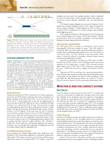
Figure 113–16. Relationship of gene structure to protein structure binding, and rapid clearance from the circulation.
in protein S. The exons, introns, mRNA, and protein structure are as
indicated. The mRNA is 2.3 kb with small 5′ and 3′ untranslated regions Gene Structure and Variations
(light blue). In the protein, Pro indicates the prepro leader sequence, The VWF gene (VWF) is located on chromosome 12p13.3, spans
GLA indicates the γ-carboxy glutamic acid (Gla) domain, T indicates approximately 180 kb, and contains 52 exons. The VWF mRNA is
199
the thrombin-sensitive region; E indicates the epidermal growth factor 8.7 kb long. There are no alternative transcripts with known biology. A
(EGF)-like domains, and the glucocorticoid hormone-binding domain
represents the sex hormone–binding globulin (SHBG)-like domain. partially inactive pseudogene that includes exons 23 to 34 is located on
199
chromosome 22p11–13. The VWF gene is very polymorphic, which
makes it sometimes difficult to distinguish between disease causing
VON WILLEBRAND FACTOR mutations and neutral gene variations.
Chapter 126 discusses the structure, function, and molecular biology of Qualitative or quantitative deficiencies in VWF cause von Wille-
VWF in detail. VWF is a large multimeric GP that is required for nor- brand disease (VWD), a mild to severe bleeding disorder. Quantitative
mal platelet adhesion to components of the vessel wall and that serves as deficiency of VWF leads to type 1 or type 3 VWD, whereas functional
a carrier for factor VIII. It is exclusively synthesized in megakaryocytes defects lead to type 2 VWD. Type 1 VWD is the most common form,
and endothelial cells and stored in specialized organelles in platelets and but type 3 VWD is the most severe. Chapter 126 discusses VWD in
endothelial cells. Release of VWF multimers from these organelles fol- detail.
lows upon a stimulus or via unstimulated basal secretion from endothe- High levels of VWF are a risk factor for venous and arterial throm-
lial cells. VWF multimers circulate at a concentration of 10 nM with bosis. Genome-wide association studies led to the identification of sev-
193
a half-life of 8 to 12 hours (see Table 113–1). Clearance of VWF multi- eral genomic loci that influence the level of VWF, including the VWF
200
mers is mainly mediated by macrophages from the liver and spleen. 194 gene itself, the ABO blood group, STXB5, and SCARA5. Polymor-
Large VWF multimers are cleaved by the plasma protease phisms in several of these loci are also associated with thrombotic risk. 201
ADAMTS-13 (a disintegrin and metalloproteinase with thrombospon-
din motifs 13). This cleavage produces the smaller size VWF multim-
195
ers that circulate in plasma. Reduced ADAMTS-13 activity is linked to FACTOR XI AND THE CONTACT SYSTEM
various microangiopathies with increased platelet activity.
FACTOR XI
Protein Structure Factor XI, which was discovered in the early 1950s, 202,203 is synthesized
The precursor protein of VWF is composed of a 22-residue signal pep- in the liver and secreted as a single-chain zymogen of 607 amino acids
tide and of a proVWF protein comprising 2791 amino acids that has 14 (Mr ≈80,000). In the circulation, factor XI is found as a homodimer
distinct domains in the order of D1-D2-D′-D3-A1-A2-A3-D4-B1-B2- (Mr ≈160,000) at a concentration of 30 nM with a plasma half-life of
B3-C1-C2-CK. Upon translocation to the ER, the signal peptide is 60 to 80 hours (see Table 113–1). All factor XI homodimers circulate in
196
cleaved off, and the proVWF dimerizes in a tail-to-tail fashion through complex with high-molecular-weight kininogen (HK). HK is thought
204
cysteines in its cysteine knot (CK) domain. During transit through to mediate binding of factor XI to negatively charged surfaces, thereby
the Golgi apparatus, proVWF dimers multimerize in a head-to-head facilitating factor XI activation. There is conflicting evidence suggest-
205
fashion through the formation of disulfide bonds between cysteine ing that HK may be also involved in the interaction of factor XI with the
residues in the D3 domain. At the same time, D1 and D2 domains activated platelet surface via GPIb. 206
are cleaved off as a single fragment to form the VWF propeptide (741
amino acids), while the remaining domains comprising 2050 amino Protein Structure
acid residues and up to 22 carbohydrate chains form mature VWF. Each factor XI subunit comprises four apple domains and a serine pro-
In the trans-Golgi network, the VWF propeptide promotes mature VWF tease domain (see Fig. 113–15). The apple domains are structured by
to assemble into high-molecular-weight multimers (Mr ≈500,000 to three disulfide bonds and form a disk-like platform on which the serine
20,000,000). These multimers subsequently aggregate into tubular protease domain rests. The dimerization of two factor XI subunits is
207
structures that are packaged into α-granules in megakaryocytes and mediated by interactions between the two apple 4 domains that involve
into Weibel-Palade bodies in endothelial cells. a disulfide bond between the Cys321 residues, hydrophobic interac-
tions, and a salt bridge, of which only the latter two are required for
von Willebrand Factor Function dimerization. The domain structure of factor XI is highly similar to
206
Upon exocytosis from Weibel-Palade bodies and at high shear rates, that of the monomer prekallikrein (PK), the zymogen of the protease
multimeric VWF unrolls from a globular to a filamental conformation kallikrein, which also circulates in complex with HK. 206
(often called VWF strings), up to many microns long, which becomes a Factor XI does not bear a Gla domain and thus does not require
high-affinity surface for the platelet GPIb–V–IX complex. Large VWF γ-carboxylation to exert its procoagulant activity. N-linked glycosylation
Kaushansky_chapter 113_p1915-1948.indd 1928 9/21/15 2:39 PM