
Page 1959 - Williams Hematology ( PDFDrive )
P. 1959
1934 Part XII: Hemostasis and Thrombosis Chapter 113: Molecular Biology and Biochemistry of the Coagulation Factors 1935
β
γ A -5.4 kb B -5.4 kb XIIIB acts as a carrier protein providing the long plasma half-life of fac-
α
-8.4 kb
tor XIII. Factor XIIIB consists of 10 Sushi domains in tandem, of which
2 4 6 8 10 2 4 8 6 4 2 the first two Sushi domains are crucial for the binding to factor XIIIA.
13 5 7 9 1 3 5 7 5 3 1
Gene 45 kb
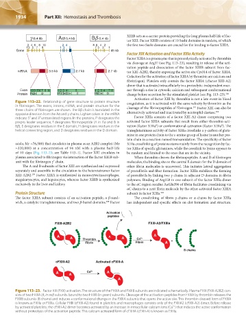
Factor XIII Activation and Factor XIIIa Activity
Factor XIIIA is a proenzyme that is proteolytically activated by thrombin
via cleavage at Arg37 (see Fig. 113–23), resulting in release of the acti-
vation peptide and dissociation of the factor XIIIB subunit from fac-
mRNA 3.5 kb 2.7 kb 2.7 kb tor XIII-A2B2, thereby exposing the active site Cys314 of factor XIIIA.
Cofactors for the activation of factor XIIIA by thrombin are calcium and
fibrin(ogen). Platelets only contain the factor XIIIA (cfactor XIII-A2)
dimer that is activated intracellularly in a proteolytic-independent man-
Exon 123567 89 1 2 3 4 5 1 2 3 4 7 8 ner through a rise in cytosolic calcium and subsequent conformational
Protein p EH D p f E H D H E p fE H D change before secretion by the stimulated platelet (see Fig. 113–23). 300
Activation of factor XIII by thrombin is not a late event in blood
Figure 113–22. Relationship of gene structure to protein structure coagulation, as it is activated with the same velocity by thrombin as the
in fibrinogen. The exons, introns, mRNA, and protein structure for the cleavage of the fibrinopeptides of fibrinogen. Factor XIII can also be
301
three chains of fibrinogen are shown. The Bβ chain is translated in the 302
opposite direction from the Aα and γ chains. Lighter colors in the mRNA alternatively activated and inactivated by neutrophil elastase.
indicate 5′ and 3′ untranslated regions. In the proteins, P designates the Factor XIIIa consists of a factor XIII-A2 dimer comprising two
prepro leader sequence, f designates fibrinopeptide (A in Aα and B in activated factor XIIIA subunits that result from either thrombin-acti-
Bβ), E designates residues in the E domain, H designates residues in the vation (factor XIIIa*) or conformational-activation (factor XIIIa°). The
helical connecting region, and D designates residues in the D domain. transglutaminase activity of factor XIIIa crosslinks a γ-carbon of gluta-
mine in one protein chain to the ε-amino group of lysine in another pro-
tein chain in a reaction named transamidation. The specificity of factor
acids; Mr ≈76,500) that circulates in plasma as an A2B2 complex (Mr XIIIa crosslinking of proteins stems mainly from the recognition by fac-
≈320,000) at a concentration of 94 nM with a plasma half-life tor XIIIa of specific glutamines, while the crosslink to lysine appears to
of 10 days (Fig. 113–23; see Table 113–1). Factor XIII circulates in be random and limited to the ones that are in the vicinity.
plasma associated to fibrinogen via interaction of the factor XIIIB sub- When thrombin cleaves the fibrinopeptides A and B of fibrinogen
unit with the fibrinogen γ′ chain. molecules, the binding site on the central E domain for the D domain of
The A and B subunits of factor XIII are synthesized and expressed other fibrin molecules is uncovered. This initiates lateral aggregation
separately and assemble in the circulation to the heterotetramer factor of protofibrils and fiber formation. Factor XIIIa stabilizes the forming
XIII-A2B2. Factor XIIIA is synthesized in monocytes/macrophages, of protofibrils by linking two γ chains in adjacent D domains in fibrin
299
megakaryocytes, and hepatocytes, whereas factor XIIIB is synthesized polymers. Binding of Arg158 in one subunit of the factor XIIIa dimer
exclusively in the liver and kidney. to the αC region residue AαGlu396 of fibrin facilitates crosslinking via
αC chains to a next fibrin molecule by the other activated factor XIIIA
Protein Structure subunit in factor XIIIa. 303
The factor XIIIA subunit consists of an activation peptide, a β-sand- The crosslinking of fibrin γ-chains or α-chains by factor XIIIa
wich, a catalytic transglutaminase, and two β-barrel domains. Factor has independent and specific effects on clot formation and structure.
298
Activation
peptides
FXIII-A2B2 FXIII-A2/FXIIIa
A A A A Ca 2+ A
Thrombin
Fibrin Fibrin A +
B chains
cFXIII-A2 Activated cFXIII-A
A A Ca 2+ A
A
Figure 113–23. Factor XIII (FXIII) activation. The structure of the FXIIIA and FXIIIB subunits are indicated schematically. Plasma FXIII (FXIII-A2B2) con-
sists of two FXIIIA (A, in red) subunits bound to two FXIIIB (in green) subunits. Cleavage of the activation peptides from FXIIIA by thrombin releases the
FXIIIB subunits (B chains) and induces a conformational change in the FXIIIA subunits that opens the active site. This thrombin cleaved form of FXIIIA
is known as FXIIIa or FXIIIa. Cellular FXIII (cFXIII-A2) found in platelets and macrophages consists only of the FXIIIA2 (cFXIII-A2) dimer. Before release
2+
by activated platelets, the cFXIII-A2 dimer becomes activated by an increase in intracellular calcium ions (Ca ) that induces the active conformation
without proteolysis of the activation peptide. This calcium activated form of cFXIIIA (cFXIII-A) is known as FXIIIa.
Kaushansky_chapter 113_p1915-1948.indd 1934 9/21/15 2:40 PM