
Page 1954 - Williams Hematology ( PDFDrive )
P. 1954
1928 Part XII: Hemostasis and Thrombosis Chapter 113: Molecular Biology and Biochemistry of the Coagulation Factors 1929
occurs at three sites in the apple 1, 2, and 4 domains (Asn82, Asn114, Deficiencies of factor XI in humans can lead to a bleeding ten-
224
Asn335) and at two sites in the serine protease domain (Asn432, dency, although not as severe as in hemophilia A or B. Deficiency of
225
Asn473). factor XI is relatively common among Ashkenazi Jews in Israel. The
human gene mutation database lists 232 mutations in the factor XI gene
Factor XI Activation and Activity (www.hgmd.org).
Activation of a factor XI subunit to factor XIa proceeds through prote- Increased levels of factor XI are a risk factor for venous throm-
olysis at Arg369 in the N-terminal region of the serine protease domain bosis. Genetic variations in the form of common single nucleotide
226
and yields two-chain activated factor XIa. There are several catalysts polymorphisms (SNPs) seem to play a role in determining the level of
capable of factor XI activation, which include the contact factor XIIa, factor XI and contribute to thrombotic risk. 227
thrombin, or factor XIa itself in the presence of negatively charged
surfaces. 208,209 However, their mechanisms differ as factor XI must be a
dimer to be activated by factor XIIa, whereas thrombin and factor XIa THE CONTACT SYSTEM: FACTOR XII,
210
lack this requirement. An activated factor XI dimer may comprise PREKALLIKREIN, AND HIGH-MOLECULAR
either one (1/2-factor XIa) or two factor XIa subunits. 211
Following activation of factor XI, binding sites for the substrate WEIGHT KININOGEN
factor IX become available in the apple 3 domain and serine protease
domain of factor XIa. 212,213 Factor XIa proteolytically activates factor IX Factor XII, HK, and PK are part of the contact system in blood coag-
to factor IXa in a calcium-dependent but phospholipid-independent ulation, which is triggered following contact activation of factor XII
manner. Both forms of the factor XIa dimer as well as monomeric factor mediated via a negatively charged surface. PK is synthesized in the liver,
XIa activate factor IX in a similar manner. 211 circulates as a zymogen, and is highly homologous to factor XI (see
Accumulating evidence supports the notion that factor XIIa– Table 113–1). Conversion into the serine protease proceeds through
dependent activation of factor XI is not essential to normal hemostasis, limited proteolysis by activated factor XII, and the generated kallikrein
but is important in pathologic thrombus formation. 214–216 Thrombin- reciprocally activates more factor XII. HK, which is also synthesized in
mediated activation of factor XI, on the other hand, seems most signifi- the liver, is a nonenzymatic cofactor that circulates in complex with fac-
cant under conditions of low tissue factor and is assumed to enhance tor XI or PK (see Table 113–1). HK is cleaved at two sites by kallikrein
clot stability through thrombin-activation of TAFI. 217,218 to release the bioactive nonapeptide bradykinin, a potent vasodilator.
Factor XI has been reported to interact with platelet GPIb, which is The contact system is at the basis of the activated partial throm-
mediated through a site within the apple 3 domain, and to platelet apo- boplastin time (APTT) assay that is widely used in clinical practice. In
lipoprotein E receptor 2 (ApoER2). 219,220 It has been proposed that the this clinical laboratory test, the negatively charged surface is provided
dimeric structure allows for simultaneous interaction with the platelet by reagents such as glass, kaolin, celite, or ellagic acid. Factor XIIa acti-
by one subunit, thereby localizing factor XI to the site of clot formation, vates factor XI, which then activates factor IX. Despite HK and PK
while binding to factor IX with the other subunit. 221 being required for a normal APTT, they appear to be dispensable for
228
Factor XIa function is regulated by the serpins protease nexin 1, coagulation in vivo. Individuals who are deficient in any of these fac-
antithrombin, C1-inhibitor, α -protease inhibitor, protein Z–dependent tors do not have a bleeding tendency, even after significant trauma or
1
protease inhibitor, and α -antiplasmin. 216,222 Platelets also contain a fac- surgery. However, factor XII, HK, and PK do participate in bacteremia
2
tor XIa inhibitor, the Kunitz-type inhibitor protease nexin 2. 223 or inflammatory responses in acute-phase reactions that do not involve
the coagulation, but the classical complement system. 228
Gene Structure and Variations
The human factor XI gene (F11) is 23 kb in length and is localized to FACTOR XII
chromosome 4q35. It consists of 15 exons and 14 introns (Fig. 113–17). Factor XII was originally reported in 1955 as the “Hageman factor,”
Each of the four apple domains is encoded by two exons. The serine named after the first identified factor XII–deficient patient. Factor
229
protease domain is encoded by five exons, with an organization similar XII is synthesized in the liver and circulates in plasma as a single-chain
to the homologous protein PK. zymogen of 596 amino acids (Mr ≈80,000) at a concentration of 500 nM
with a half-life of 50 to 70 hours (see Table 113–1).
1 2 3 4 56 78 9101112131415
Gene 23 kb Protein Structure
Factor XII, which is homologous to plasminogen activators, consists
of an N-terminal fibronectin type I domain, an EGF-like domain,
a fibronectin type II domain, a second EGF-like domain, a kringle
domain, a proline-rich region, and a C-terminal serine protease domain
mRNA 2.1 kb (see Fig. 113–15). The proline-rich region is unique to factor XII, as it is
not found in any of the other serine proteases.
Factor XII comprises an O-linked fucose in EGF 1 (Thr90),
N-linked glycosylation sites in the kringle domain (Asn230) and the
Exon 2 34 56 7 8 9 10 11 12 13 14 15 serine protease domain (Asn414), and several O-linked glycosylation
Protein ProA1 A2 A3 A4 Catalytic domain sites in the kringle domain and proline-rich region. 230,231
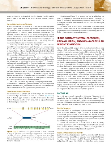
Figure 113–17. Relationship of gene structure to protein structure
in factor XI. The exons, introns, mRNA, and protein structure are as indi- Factor XII Activation and Activity
cated. The mRNA is 2.1 kb with small 5′ and 3′ untranslated regions (light Limited proteolysis by kallikrein at Arg353 in factor XII yields the acti-
blue). In the protein, Pro indicates the prepro leader sequence, A1 to vated two-chain α-factor XIIa, in which the heavy chain (the fibronec-
A4 indicate apple domains 1 to 4, and the serine protease (catalytic) tin types I and II domains, both EGF domains, the kringle domain,
domain is indicated. and proline-rich region; Mr ≈52,000) and light chain (serine protease
Kaushansky_chapter 113_p1915-1948.indd 1929 9/21/15 2:40 PM