
Page 1958 - Williams Hematology ( PDFDrive )
P. 1958
1932 Part XII: Hemostasis and Thrombosis Chapter 113: Molecular Biology and Biochemistry of the Coagulation Factors 1933
During fibrin monomer polymerization, other plasma proteins
D E D also bind to the surface of the developing meshwork. These include ele-
ments of the fibrinolytic system and a variety of adhesive proteins, such
as fibronectin, thrombospondin, and VWF. These surface proteins influ-
ence the generation, crosslinking, and lysis of fibrin. Fibrin(ogen) also
has specific integrin-binding sites that are essential for platelet binding.
The thrombin that initiates fibrin polymerization also activates factor
XIII, which stabilizes the fibrin polymer by crosslinking. Factor XIIIa
also crosslinks other bound proteins, for example, PAI-1, vitronectin,
fibronectin, and α -antiplasmin, to the fibrin network.
2
Figure 113–20. Structure of fibrinogen. Fibrinogen is a dimer. Each Once formed, the fibrin mesh can be degraded by the fibrinolytic
monomer consists of three chains: Aα shown in light blue, Bβ shown in system. Plasmin cleaves fibrin and fibrinogen in an ordered sequence
pink, and γ shown in dark blue. The disulfides that link the two monomers at arginyl and lysyl bonds, giving rise to a series of soluble degrada-
are in the central E domain. The D domains consist primarily of the C-ter- tion products. In this process, the crosslink between two D fragments
296
minal regions of the Bβ and γ chains. The helical region connecting the remains intact, resulting in the formation of a fragment consisting of
two domains consists of all three chains intertwined. (Reproduced with per- two D domains and one E domain, called D-dimer. Circulating
mission from Côté HC, Lord ST, Pratt KP: Gamma-Chain dysfibrinogenemias: D-dimer concentrations are often measured as a surrogate marker of
Molecular structure-function relationships of naturally occurring mutations activated coagulation.
in the gamma chain of human fibrinogen. Blood 92(7):2195–2212, 1998.)
In addition to its obvious procoagulant role in stabilizing the initial
platelet hemostatic plug, fibrin can also act as an important inhibitor of
Because human fibrinogen is subject to modification at a number of thrombin generation. Fibrin functions as “antithrombin I” by seques-
different sites both during and after biosynthesis, the fibrinogen present tering thrombin in the developing fibrin clot, and also by reducing the
in the circulation is a heterogeneous mixture of molecules. These nor- catalytic activity of fibrin-bound thrombin. 297
mal variants are caused by alternative splicing, modification of certain
amino acids by sulfation, phosphorylation, and hydroxylation, different Gene Structure and Variations
degrees of glycosylation, and proteolysis. It has been estimated that the The genes for the three chains of fibrinogen are found within a 50-kb
number of nonidentical fibrinogen molecules that can be produced by region on chromosome 4 at q23-q32 (Fig. 113–22). The genomic
these mechanisms is in excess of 1 million. Some of these variations sequences show a high degree of homology, suggesting they were
289
may have significant functional consequences. For example, the level of derived through duplication of a common ancestral gene. The homol-
one variant of fibrinogen with an alternatively spliced γ chain (fibrino- ogy extends to sites upstream of the gene, suggesting that common reg-
gen-γ′) is associated with a risk of venous thrombosis. 290 ulatory elements may reside in these areas, thus helping to coordinate
synthesis of the three chains.
Fibrinogen Activation and Fibrin Function The physiologic importance of fibrinogen is underscored by the
Thrombin binds to the central domain of fibrinogen and proteolytically bleeding diathesis associated with afibrinogenemia and some dysfibrin-
releases two fibrinopeptides A (Aα, residues 1 to 16) and two fibrinopep- ogenemias (Chap. 125). Other dysfibrinogenemias are associated with
tides B (Bβ, residues 1 to 14) from each fibrinogen molecule. Release thromboembolic disease. Although afibrinogenemia is associated with
291
of the fibrinopeptides exposes binding sites in the E domain that have a bleeding tendency, it is usually not as severe as classical hemophilia.
complementary sites in the D domains of other fibrin monomers. 292,293
These complementary binding sites lead to the initial formation of
two-stranded protofibrils with a half-staggered overlap configuration FACTOR XIII
(Fig. 113–21). Protofibrils then aggregate into thick fibers that branch The GP factor XIII is a protransglutaminase that, upon activation,
into a meshwork of interconnected thick fibers. The half-staggered crosslinks and stabilizes fibrin clots. Plasma factor XIII is a het-
294
298
overlap of the fibrin monomers gives a characteristic cross-banded pat- erotetramer consisting of two factor XIIIA subunits (731 amino
tern on electron micrographs. 295 acids; Mr ≈83,000) bound to two factor XIIIB subunits (641 amino
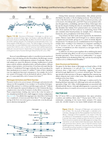
Fibrinogen Figure 113–21. Cleavage of fibrinogen and polymeriza-
E D
tion of fibrin. The structure of fibrinogen is indicated sche-
a a matically. Cleavage sites for fibrinopeptide A by thrombin are
Thrombin
Fibrin monomer shown. Cleavage of the B peptide is not shown in this fig-
ure. Release of fibrinopeptide A exposes binding sites in the
a a
AA E domain that match complementary sites in the D domain.
Protofibril Fibrin monomers polymerize by half-staggered overlaps. Poly-
merization can also lead to branched structures. (Reproduced
with permission from Côté HC, Lord ST, Pratt KP: Gamma-Chain
dysfibrinogenemias: Molecular structure-function relationships
of naturally occurring mutations in the gamma chain of human
fibrinogen. Blood 92(7):2195–2212, 1998.)
Branching
Three-dimensional thickening
of the fibrils into fibers
Kaushansky_chapter 113_p1915-1948.indd 1933 9/21/15 2:40 PM