
Page 761 - Williams Hematology ( PDFDrive )
P. 761
736 Part VI: The Erythrocyte Chapter 48: The Thalassemias: Disorders of Globin Synthesis 737
0
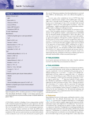
TABLE 48–4. Hereditary Persistence of Fetal Hemoglobin that carry β -thalassemia mutations, the clinical phenotype is converted
from HPFH to δβ-thalassemia, albeit with different hemoglobin A
Deletion (Pancellular ) * levels. 2
(δβ) 0 In some cases, other nondeletional forms of HPFH have been
related to small structural changes in the β-globin gene cluster (see
Black (HPFH 1)
Table 48–4). Although strictly speaking not a true form of HPFH,
Ghanaian (HPFH 2) because even in homozygotes it may not be associated with increased
Indian (HPFH 3) hemoglobin F levels, the T→C polymorphism at position –158 to the
121
G γ-globin gene might be associated with an increased output of hemo-
Italian (HPFH 4 and 5)
globin F under conditions of erythropoietic stress.
Vietnamese (HPFH 6) Other forms of HPFH are characterized by the persistence of low
+
A
G γ ( γ β) (Hgb Kenya) levels of fetal hemoglobin production distributed in a heterocellular
manner. In all populations studied, a small proportion of individuals
Nondeletion
have an increased amount of hemoglobin F and F cells, that is, red
Linked to β-globin gene cluster (pancellular ) cells that can be detected when blood films are treated with antibod-
*
G γ β + ies against hemoglobin F. Although this condition originally was called
the Swiss form of HPFH because it was first recognized in Swiss army
Black γ-202 C→G
G
122
recruits, it is observed in every racial group. Using a variety of genetic
G
Tunisian γ-200+C approaches, it has become clear that a number of genes may be involved
G
Black/Sardinian γ-175 T→C in the generation of heterocellular HPFH, including loci at Xp22.2-
p22.3,6q23,8q, and 2p15 123–128 ; the latter linkage has been identified as
G
Japanese γ-114 C→T
the oncogene BCL11A. The mechanism whereby these different loci
G
Australian γ-114 C→G affect the level of F cells in normal individuals and increase their lev-
A γ β + els in conditions like thalassemia and sickle cell anemia remain to be
determined, but their coinheritance with these conditions may have an
Greek/Sardinian/Black γ-117 G→A extremely beneficial effect of their associated phenotypes. 129
A
British γ-198 T→C
A
Black γ-202 C→T δ-THALASSEMIA
A
Italian/Chinese γ-196 C→T Several point mutations and deletions that reduce δ-globin synthesis
A
have been described. They are summarized in reference 7.
Brazilian γ-195 C→G
A
Black γ-175 T→C α-THALASSEMIA
A
Black γ-114 to –102 (del)
A
Table 48–5 summarizes the different classes of α-thalassemia mutations.
Georgia γ-114 C→T The α-globin gene haplotype can be written αα, indicating the α and
A
1
A
G γ γ β + α genes, respectively. A normal individual has the genotype αα/αα. A
2
deletion involving one (–α) or both (– –) α genes can be further clas-
Linked to β-globin gene cluster (heterocellular )
*
sified based on its size, written as a superscript; thus, –α indicates a
3.7
Atlanta deletion of 3.7 kb including one α gene. When the sizes of the deletions
Czech are not established, a superscript describing their geographic or fam-
ily origin is useful; thus, – – MED describes a deletion of both α genes
Seattle
first identified in individuals of Mediterranean origin. In thalassemia
G
Others (including some cases of γ-158 T→C) haplotypes in which both genes are intact, that is, nondeletion lesions,
ND
Unlinked to β-globin gene cluster (heterocellular ) * the nomenclature α α is given, with the superscript ND indicating
the gene is thalassemic. However, when the precise molecular defect
Chromosome 6 is known, as in hemoglobin Constant Spring, for example, α α can be
ND
Others replaced by the more informative α α. The molecular pathology and
CS
population genetics of the α-thalassemias have been the subject of sev-
Hgb, hemoglobin; HPFH, hereditary persistence of fetal hemoglobin. eral extensive reviews. 7,41,45,130,131
*The intercellular distribution of Hgb F is not always reported, and
some inconsistencies are present within groups. Complete details are α -Thalassemia
0
given in Ref. 7. Many deletions that involve both α genes, and therefore abolish α-chain
production from the affected chromosome, have been described
(Fig. 48–10). Several of the 3′ breakpoints fall within a 6- to 8-kb region
7
of DNA likely is involved in binding of trans-acting proteins involved at the 3′ end of the α-globin complex, suggesting this represents a break-
132
in the normal developmental repression of γ-gene expression, either by point cluster region with a high level of recombination. In at least five
decreasing the affinity for an inhibitory factor normally present in adult of the deletions, the 5′ breakpoints also appear to cluster. This gives rise
life or by increasing the affinity for a factor promoting gene expression. to a situation in which the 5′ breakpoints are located approximately the
The most common of these conditions are Greek γβ HPFH and a same distance apart and in the same order along a chromosome as their
+
A
form of γβ HPFH, which has been found in several different African respective 3′ breakpoints. It is possible that such staggered deletions
+
G
populations. If the upstream point mutations associated with persistent arise from illegitimate recombination events that delete an integral num-
γ-chain production occur on the same chromosome as β-globin genes ber of chromatin loops as they pass through their nuclear attachment
Kaushansky_chapter 48_p0725-0758.indd 736 9/18/15 2:57 PM