
Page 1004 - Clinical Immunology_ Principles and Practice ( PDFDrive )
P. 1004
968 Part Seven Organ-Specific Inflammatory Disease
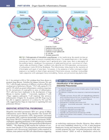
Steady state Initiation of injury and repair Dysregulated repair
Alveolus
Type I epithelial cell
Monocytes
Dendritic cell Alveolar
macrophages
Lymphocytes Fibrosis
Type II epithelial cell
PMNs
ECM ECM
Resolution
Fibroblasts Fibroblasts
1. Denudation of barrier
2. Cytokine/chemokine expression
3. Recruitment of inflammatory cells
4. Proliferation of type II epithelial cells
5. DC maturation
6. Fibroblasts express ECM
FIG 72.1 Pathogenesis of Interstitial Lung Disease. In the healthy lung, the alveoli maintain an
antiinflammatory state to prevent unwanted inflammation. The predominant cells in the healthy
alveolus are macrophages and types I and II epithelial cells. Upon injury, there is denudation of
the epithelial barrier, resulting in the expression of cytokines/chemokines, recruitment of inflam-
matory cells, proliferation of type II epithelial cells, dendritic cell (DC) maturation, and expression
of extracellular matrix (ECM) by fibroblasts. This inflammatory milieu leads to the resolution of
inflammation and repair of lung injury. Conversely, in the presence of either prolonged antigen
exposure or an inability to clear antigen, persistent inflammation ensues, resulting in extracellular
matrix deposition with subsequent tissue remodeling, fibrosis, and permanent lung dysfunction.
IL-13. In contrast to IFN-γ, Th2 cytokines have been shown to KeY COnCePtS
promote lung fibrosis. Therefore the balance between Th1 and
Th2 T cells through expression of different cytokines affects the Classification of the Idiopathic
10
development of pulmonary fibrosis. Th17 cells express IL-17A Interstitial Pneumonias
and IL-17F, which are potent inflammatory cytokines important • The idiopathic interstitial pneumonias comprise a group of eight histologi-
for the recruitment of neutrophils to areas of inflammation. cally distinct disorders.
Th17 cells have been implicated in the development of lung • The distinction is important since response to treatment and prognosis
fibrosis in murine models. 11,12 Conversely, regulatory T cells differs:
(Tregs) suppress pathogenic T-cell responses that promote • Idiopathic pulmonary fibrosis (IPF) with histopathology labeled as
inflammation (Chapter 18). In patients with IPF, Tregs may be usual interstitial pneumonitis (UIP): the pathology of UIP can occur
in other diseases such as connective tissue diseases
less able to suppress expression of cytokines by Th1 and Th2 • Acute interstitial pneumonitis (AIP)
cells, suggesting that Tregs are important in regulating ILD and • Desquamative interstitial pneumonitis (DIP)
pulmonary fibrosis. 13 • Respiratory bronchiolitis–associated interstitial lung disease
(RB-ILD)
IDIOPATHIC INTERSTITIAL PNEUMONIAS • Nonspecific interstitial pneumonia (NSIP)
• Lymphocytic interstitial pneumonia (LIP)
• Cryptogenic organizing pneumonia (COP)
Idiopathic interstitial pneumonias (IIPs) are a group of diffuse
• Idiopathic pleuroparenchymal fibroelastosis (IPPFE)
inflammatory and/or fibrotic lung disorders that include IPF, • To differentiate between these diseases, a thoracoscopic lung biopsy
acute interstitial pneumonitis (AIP), desquamative interstitial is often required.
pneumonitis (DIP), respiratory bronchiolitis–associated interstitial
lung disease (RB-ILD), nonspecific interstitial pneumonia (NSIP),
cryptogenic organizing pneumonia (COP), lymphocytic interstitial
pneumonia (LIP), and idiopathic pleuroparenchymal fibroelastosis an underlying autoimmune disorder. However, these subjects
14
(IPPFE). The diagnosis of IIPs requires the exclusion of con- do not meet established criteria for a CTD. A joint European
nective tissue diseases (CTDs), drug toxicity, and environmental Respiratory Society and American Thoracic Society Task Force
exposures, and a thoracoscopic lung biopsy is often required. In recently proposed the term “interstitial pneumonia with auto-
addition, some patients with IIPs have clinical features suggesting immune features” (IPAF) as well as classification criteria for