
Page 858 - Clinical Immunology_ Principles and Practice ( PDFDrive )
P. 858
CHaPTEr 60 Systemic Autoinflammatory Syndromes 829
There are two exceptional TNFRSF1A mutations: R92Q and
Tumor Necrosis Factor Receptor–Associated P46L. These mutations do not lead to receptor misfolding and
Periodic Syndrome are present in low frequency in the general population. They
Mutations in the gene TNFRSF1A are responsible for TRAPS. This may, however, cause a mild inflammatory phenotype.
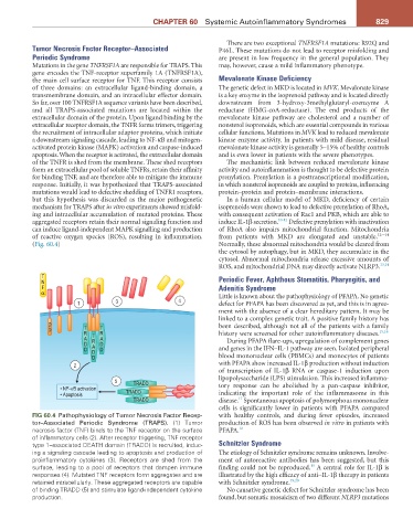
gene encodes the TNF-receptor superfamily 1A (TNFRSF1A),
the main cell surface receptor for TNF. This receptor consists Mevalonate Kinase Deficiency
of three domains: an extracellular ligand-binding domain, a The genetic defect in MKD is located in MVK. Mevalonate kinase
transmembrane domain, and an intracellular effector domain. is a key enzyme in the isoprenoid pathway and is located directly
So far, over 100 TNFRSF1A sequence variants have been described, downstream from 3-hydroxy-3methylglutaryl-coenzyme A
and all TRAPS-associated mutations are located within the reductase (HMG-coA-reductase). The end products of the
extracellular domain of the protein. Upon ligand binding by the mevalonate kinase pathway are cholesterol and a number of
extracellular receptor domain, the TNFR forms trimers, triggering nonsterol isoprenoids, which are essential compounds in various
the recruitment of intracellular adaptor proteins, which initiate cellular functions. Mutations in MVK lead to reduced mevalonate
a downstream signaling cascade, leading to NF-κB and mitogen- kinase enzyme activity. In patients with mild disease, residual
activated protein kinase (MAPK) activation and caspase-induced mevalonate kinase activity is generally 5–15% of healthy controls
apoptosis. When the receptor is activated, the extracellular domain and is even lower in patients with the severe phenotypes.
of the TNFR is shed from the membrane. These shed receptors The mechanistic link between reduced mevalonate kinase
form an extracellular pool of soluble TNFRs, retain their affinity activity and autoinflammation is thought to be defective protein
for binding TNF, and are therefore able to mitigate the immune prenylation. Prenylation is a posttranscriptional modification,
response. Initially, it was hypothesized that TRAPS-associated in which nonsterol isoprenoids are coupled to proteins, influencing
mutations would lead to defective shedding of TNFR1 receptors, protein–protein and protein–membrane interactions.
but this hypothesis was discarded as the major pathogenetic In a human cellular model of MKD, deficiency of certain
mechanism for TRAPS after in vitro experiments showed misfold- isoprenoids were shown to lead to defective prenylation of RhoA,
ing and intracellular accumulation of mutated proteins. These with consequent activation of Rac1 and PKB, which are able to
aggregated receptors retain their normal signaling function and induce IL-1β secretion. 10-12 Defective prenylation with inactivation
can induce ligand-independent MAPK signalling and production of RhoA also impairs mitochondrial function. Mitochondria
of reactive oxygen species (ROS), resulting in inflammation. from patients with MKD are elongated and unstable. 12-–14
(Fig. 60.4) Normally, these abnormal mitochondria would be cleared from
the cytosol by autophagy, but in MKD, they accumulate in the
cytosol. Abnormal mitochondria release excessive amounts of
ROS, and mitochondrial DNA may directly activate NLRP3. 13,14
T
N Periodic Fever, Aphthous Stomatitis, Pharyngitis, and
F Adenitis Syndrome
α Little is known about the pathophysiology of PFAPA. No genetic
1 3 4 defect for PFAPA has been discovered as yet, and this is in agree-
ment with the absence of a clear hereditary pattern. It may be
linked to a complex genetic trait. A positive family history has
D been described, although not all of the patients with a family
D T T 15,16
R T R history were screened for other autoinflammatory diseases.
A R A During PFAPA flare-ups, upregulation of complement genes
D A D
D D D and genes in the IFN–IL-1 pathway are seen. Isolated peripheral
D blood mononuclear cells (PBMCs) and monocytes of patients
2 with PFAPA show increased IL-1β production without induction
of transcription of IL-1β RNA or caspase-1 induction upon
5 TRADD lipopolysaccharide (LPS) stimulation. This increased inflamma-
• NF-κB activation tory response can be abolished by a pan-caspase inhibitor,
• Apoptosis TRADD indicating the important role of the inflammasome in this
17
TRADD disease. Spontaneous apoptosis of polymorphous mononuclear
cells is significantly lower in patients with PFAPA compared
FIG 60.4 Pathophysiology of Tumor Necrosis Factor Recep- with healthy controls, and during fever episodes, increased
tor–Associated Periodic Syndrome (TRAPS). (1) Tumor production of ROS has been observed in vitro in patients with
necrosis factor (TNF) binds to the TNF receptor on the surface PFAPA. 18
of inflammatory cells (2). After receptor triggering, TNF receptor
type 1–associated DEATH domain (TRADD) is recruited, induc- Schnitzler Syndrome
ing a signaling cascade leading to apoptosis and production of The etiology of Schnitzler syndrome remains unknown. Involve-
proinflammatory cytokines (3). Receptors are shed from the ment of autoreactive antibodies has been suggested, but this
surface, leading to a pool of receptors that dampen immune finding could not be reproduced. A central role for IL-1β is
19
responses (4). Mutated TNF receptors form aggregates and are illustrated by the high efficacy of anti–IL-1β therapy in patients
retained intracellularly. These aggregated receptors are capable with Schnitzler syndrome. 19,20
of binding TRADD (5) and stimulate ligand-independent cytokine No causative genetic defect for Schnitzler syndrome has been
production. found, but somatic mosaicism of two different NLRP3 mutations