
Page 1576 - Hall et al (2015) Principles of Critical Care-McGraw-Hill
P. 1576
CHAPTER 115: The Transplant Patient 1095
Endobronchial surfactant: The use of exogenous surfactant has been score (between 0 and 99—based on the percent of the population that
shown to reduce PGD in animal models and in some preliminary recipient’s blood would react with) is assigned. However, controversy
studies in humans. Further research is underway to determine if exists surrounding how to manage the patient with an elevated PRA. In
surfactant has any effect on patient-relevant outcomes. 52-55 the Toronto Lung Transplant program, if a patient has a positive PRA,
virtual donor–specific antibody test, as well as positive cross match, we
Acute Rejection: The incidence of hyperacute rejection, acute rejec- proceed with the administration of plasma exchange intraoperatively
tion, and chronic rejection has declined since the improvement in and on days 1, 2, 3, and 5 followed by administration of intravenous
immunosuppressive regimens. Hyperacute rejection is currently a immunoglobulin and MMF (in lieu of azathioprine) as consolidation
rare complication due to improvements in detecting HLA antibodies. therapy. Antithymocyte globulin follows intravenous immunoglobulin
Thirty six percent of patients will experience at least one episode in the absence of evidence of infection.
of rejection. While chronic rejection could cause complications at
31
a later time in the life of a transplant recipient, it is not commonly Bleeding: One of the most common complications requiring a return to
seen in the ICU as the primary etiology for presentation; however, the OR is postoperative bleeding. An unexplained drop in hemoglobin
beyond the first year of transplant, chronic rejection accounts for even in the absence of blood loss through thoracostomy tube drain-
25% of deaths. 56 age should raise this as a consideration. Fortunately, it has not been
Hyperacute Rejection Hyperacute rejection occurs when the recipient associated with an increase in perioperative mortality. Risk factors for
has preformed antibodies to antigens on donor tissue (alloantigens). postoperative hemorrhage include the presence of pleural adhesions
Fulminant rejection occurs within minutes of reperfusion. It manifests in the recipient (more commonly seen in patients with cystic fibrosis,
as acute respiratory failure, gross pulmonary edema with airway hemor- eosinophilic granuloma, or lymphangioleiomyomatosis) and the use of
rhage, and thrombosis within the graft. With improvement in screen- cardiopulmonary bypass.
ing techniques for preformed antihuman leukocyte antigen antibodies Airway Complications: Six different airway complications of post-lung
before transplant and the use of plasma exchange and thymoglobulin in transplant include bronchial dehiscence, endobronchial infections, exo-
a highly sensitized patient, the incidence has decreased. It is uncommon phytic excessive granulation tissue formation, tracheobronchomalacia,
as it is only encountered when there is ABO incompatibility or if the bronchial fistulas, and bronchial stenosis. Bronchial dehiscence and
recipient is sensitized to alloantigens from previous blood transfusions. endobronchial infections can arise in the immediate posttransplant
Management includes urgent plasma exchange followed by agents to period and will be the focus of this section. Table 115-7 provides more
modify B-cell response such as rituximab. 57 detailed information on the airway complications that may challenge the
Acute Rejection Acute rejection is commonly seen after the first week to the management of patients postoperatively. Bronchial complications were
first year posttransplant with the highest prevalence in the first 100 days a significant source of morbidity and mortality in the early era of trans-
after transplant. It is mediated by a recipient T-cell response against the plant being present in up to 80% of patients; however, with improved
graft. It is uncommon that an isolated episode of acute rejection would surgical techniques and newer immunosuppressive agents, they now
require ICU care; however, it has the potential to complicate the wean- occur in only 7% to 18% of patients. 58
ing process of a newly transplanted patient as it could develop early Bronchial necrosis and dehiscence is a potentially devastating conse-
after transplant. Perivascular and interstitial infiltration of mononuclear quence of transplant and is associated with a high mortality. Ischemia of
cells are found on transbronchial or open lung biopsy. Clinically, acute the donor bronchus in the immediate posttransplant period is often the
rejection manifests as perihilar infiltrates, leukocytosis, hypoxia, airway etiology of bronchial dehiscence. The dual blood supply to the bronchus
inflammation, and low-grade fever in the absence of cultured organisms. in normal lungs arises from the pulmonary artery and from bronchial
Treatment includes pulse steroids (methylprednisolone 10-15 mg/kg) for arteries originating from the intercostals off of the descending aorta.
3 to 5 days followed by a 2- to 3-week steroid taper to the usual dose. The bronchial arterial circulation to the anastomosis is disrupted during
Patients noted to have a positive panel of reactive antibodies (PRA) surgery and not reestablished until 2 to 4 weeks posttransplant. Therefore,
are considered sensitized and are at greater risk of acute rejection post- the new lung’s airway anastomosis is dependent on the relatively low
transplant. PRAs screen for antihuman leukocyte antibodies, and a pressured, poorly oxygenated collateral blood flow from the pulmonary
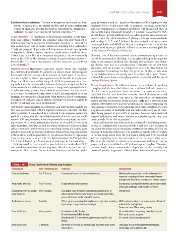
TABLE 115-7 Airway Complications Following Lung Transplant
Complication Time to Presentation Definition Management
Bronchial dehiscence First 1-5 weeks Ischemia and necrosis of bronchial anastomosis Minimize airway pressures, consider antimicrobials if
suspected coexisting infection, conservative/invasive
management depends on severity as per thoracic surgeon
Anastomotic infections First 1-5 weeks Fungal Infections of anastomosis Bronchoscopic draining/debridement; systemic/aerosolized
antifungals; antifungal prophylaxis for prevention
Exophytic excessive granulation Weeks-months Granulation tissue formation secondary to overstimulation of Debridement
tissue formation inflammatory mediators; ischemia and subsequent remodelling
process of stenosis. Similar to keloid formation
Tracheobronchomalacia 4 months 50% or greater narrowing of airway lumen on expiration secondary Mild: treat concomitant process causing exacerbation of
to pathologic changes in airway cartilages malacia (infection/rejection)
Severe: nocturnal PPV or airway stenting
Bronchial fistulae Weeks-months Bronchopleural (BP) fistulae BP: antimicrobials, thoracotomy tube, fistula closure
Bronchomediastinal (BM) fistulae BM: consult thoracic surgeon
Bronchovascular (BV): bronchoaortic/pulmonary artery/left atrial BV: consult thoracic/vascular surgeon
fistulae
Bronchial stenosis 2-9 months Narrowing of bronchus usually at surgical anastomotic site (some Endoscopic management (balloon bronchoplasty,
occur distally) cryotherapy, stent placement, etc)
section10.indd 1095 1/20/2015 9:19:55 AM