
Page 1577 - Hall et al (2015) Principles of Critical Care-McGraw-Hill
P. 1577
1096 PART 10: The Surgical Patient
artery. Hypotension, compression from high airway pressures, and reper- Infectious Complications in this chapter). Computed tomographic
fusion injury leading to airway edema may also compromise flow and (CT) scanning of the chest and bronchoscopic sampling of the distal
can contribute to ischemia. Ultimately this can lead to an environment airways may assist in ruling out an infectious cause. A trial of diuresis,
for infection, extensive necrosis and then dehiscence. Late complications echocardiogram, or placement of a pulmonary arterial catheter may
of stenosis can arise as healing and remodeling takes place. Necrosis and be required to evaluate the contribution of volume overload to the
dehiscence is suspected in the setting of persistent air leak, lung collapse, opacities. The role of lung biopsy in establishing the cause of radio-
difficulty weaning from the ventilator, subcutaneous emphysema, pneu- graphic worsening is controversial. Transbronchial lung biopsy may
momediastinum, and pneumothorax. The anastomosis must be carefully lack sufficient sensitivity in the perioperative setting. However, the
60
examined with flexible bronchoscopy. In addition, dehiscence may be utility of open lung biopsy (OLB) is variable, 61,62 and hence it should be
further complicated by peribronchial abscess formation; therefore, a CT carefully considered if the etiology of the lung infiltrates remains uncer-
scan to rule out mediastinal air or surrounding infection/abscess should tain. In one retrospective series of 48 open biopsies from 42 patients,
also be performed. Management includes minimizing high airway 32 (67%) of the biopsies confirmed the clinicians’ initial suspicions and
pressures, initiating appropriate antimicrobials if suspicion of secondary prompted the initiation of “new therapy” in 30 (71%) of the patients.
61
infection exists and either conservative management, endobronchial A new diagnosis was made following 14 of the biopsies (29%). Only
repair, or open repair depending on the severity. two patients (4% of all OLBs) had a nondiagnostic OLB. Four biopsies
Endobronchial infections occur due to the impairment of regional (8% of all OLBs), including the two nondiagnostic OLBs, did not result
defense mechanisms (from ischemia, decreased mucociliary clearance, in any change in therapy. Complications of the procedure were rare,
minimized cough reflex) and influenced by the ongoing use of high- though three (7%) patients developed an air leak, which persisted more
dose immunosuppressant medications. This regional and systemic effect than 7 days. In contrast, an earlier report of 38 biopsies (representing
on immunological integrity fosters a rich environment for bacterial 32 patients) found that early open lung biopsies (performed <45 days
and fungal overgrowth. Saprophytic infections are the most frequently after transplant) were not useful, and resulted in a change in therapy for
seen organisms as they are airborne and maintain nourishment from only 1 of 11 cases. 62
nonliving organic material in ischemic and necrotic debris. Aspergillus
is the most frequently seen organism. Treatment includes a combination Long-Term Complications/Chronic Lung Allograft Dysfunction: Chronic
of bronchoscopic drainage, debridement, and systemic as well as inhaled lung allograft dysfunction (CLAD) is a long-term complication and the
antifungals. Antifungal prophylaxis has been advocated to minimize the forme fruste of chronic rejection in the transplant recipient. Previously,
risk of fungal anastomotic infections and is used in greater than 70% of chronic rejection was termed as bronchiolitis obliterans syndrome
the transplant programs within 24 hours after the procedure. 58 (BOS) which was characterized by obliterative bronchiolitis resulting
Arrhythmias: Atrial fibrillation is seen in 20% of patients in the postop- in a progressive fall in FEV over time that was not attributed to acute
1
rejection, infection of mechanical obstruction. However, in recent
erative period with the peak incidence at 2 to 4 days. For most patients years, an additional form of chronic rejection was identified char-
59
(93%), the arrhythmia is isolated to the postoperative period and most acterized by fibrosis in peripheral tissues and restrictive physiology,
revert back to normal sinus rhythm before discharge. Risk factors for termed restrictive allograft syndrome (RAS). Additional long-term
atrial fibrillation include older age, IPAH, and extremes of weight. There complications are outlined in Table 115-9. In one series that followed
is considerable debate regarding the management of atrial fibrillation in patients over a 10-year period 74% of patients had developed BOS.
43
the postoperative period and no consensus has been established on the Chronic rejection remains one of the most common causes of death
optimal management. Rationale for cardioversion follows the reasoning and disability in the posttransplant period. Causes for CLAD are not
that patients are often refractory to rate control attempts and that the completely understood but believed to be related to both alloimmune
short cardiac filling time in the immediate postoperative period leads to and nonimmune mechanisms. Risk factors include more frequent epi-
further pulmonary congestion which is poorly tolerated in these patients. sodes of acute rejection, gastroesophageal reflux, and CMV infection.
However, there are no clinical data to support this notion. Furthermore, Clinically BOS patients have predominantly a progressive obstructive
concern about amiodarone-induced lung toxicity may restrict therapeutic disease pattern and RAS patients have predominantly a restrictive
choices. Unless poorly tolerated, rate control may be safely and effectively lung disease pattern that could progress to end-stage lung disease and
pursued with β-blockers, calcium-channel blockers, or digoxin. be exacerbated by infections prompting bouts of acute respiratory
New Airspace Opacities in the Perioperative Period: Defining the etiol- failure. Management includes the use of bronchodilators, corticoste-
ogy of new or progressive airspace opacities in the perioperative lung roids, aggressive treatment of reflux, and modification of immuno-
transplant period is a frequent dilemma. The differential diagnosis suppressive agents. Ventilatory management of these patients may be
for new airspace disease is outlined in Table 115-8 and it may repre- challenging but follows from principles used to manage patients with
sent PGD, volume overload, early rejection, or infection (see section other forms of obstructive or restrictive lung disease.
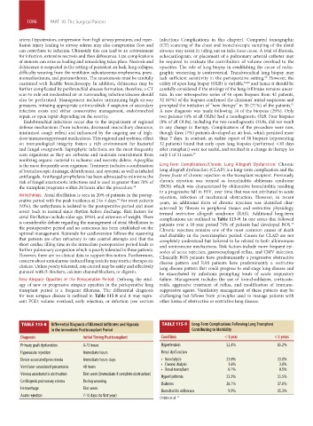
TABLE 115-8 Differential Diagnosis of Bilateral infiltrates and Hypoxia TABLE 115-9 Long-Term Complications Following Lung Transplant
in the immediate Posttransplant Period Contributing to Morbidity
Diagnosis initial Timing Posttransplant Condition <1 year <5 years
Primary graft dysfunction 0-72 hours Hypertension 52.4% 85.2%
Hyperacute rejection Immediate hours Renal dysfunction
Donor-associated pneumonia Immediate hours-days • Nondialysis 23.0% 33.0%
• Chronic dialysis 1.6% 3.0%
Ventilator-associated pneumonia 48 hours
• Renal transplant 0.1% 0.5%
Venous anastomotic obstruction First week (immediate if complete obstruction)
Hyperlipidemia 23.3% 55.5%
Cardiogenic pulmonary edema During weaning
Diabetes 26.1% 37.0%
Hemorrhage First week
Bronchiolitis obliterans 9.5% 35.3%
Acute rejection 7-10 days (to first year)
Christie et al. 31
section10.indd 1096 1/20/2015 9:19:55 AM