
Page 1814 - Hall et al (2015) Principles of Critical Care-McGraw-Hill
P. 1814
CHAPTER 129: Dermatologic Conditions 1283
cardiac arrhythmias, urticaria, and pruritus. Drug-induced anaphylaxis
TABLE 129-2 Drug Eruption Checklist: Information to Be Elicited From the
Patient’s Medical Records or Family occurs in 1 of 2700 hospitalized patients and is most frequently induced
by β-lactam antibiotics (including penicillin), radiocontrast medium,
(1) Time of onset and course of the reaction intravenous anesthetic drugs, aspirin, other NSAIDs, and opiates. In
(2) Dosage and time of initiation or discontinuation of any medications, including over- the United States, the most common cause of anaphylaxis is penicillin.
9
the-counter and alternative products Treatment of urticaria consists of discontinuation of the offending drug
(3) The patient’s previous exposure to this or other related medications and administration of oral antihistamine H blockers. These include
1
(4) Any previous history of adverse drug reaction (ADR), its management, and any diphenhydramine, hydroxyzine, and the nonsedating agents, loratadine,
measures taken to prevent future ADRs cetirizine, and fexofenadine. If anaphylaxis develops, emergency treatment
(5) The patient’s medical problems is instituted with intramuscular or subcutaneous epinephrine, high-flow
(6) Any physical or laboratory abnormalities present with the ADR, with special attention oxygen and airway management, intravenous diphenhydramine, ste-
to organ systems involved roids, fluids, vasopressors, and cardiopulmonary resuscitation, as needed
(Chap. 128). Skin testing with the offending agent is usually positive in
IgE-mediated reactions.
TABLE 129-3 Indicators that an Adverse Drug Reaction May Become Serious Cytotoxic (type II) reactions are mediated by IgG and complement,
Cutaneous Findings Systemic Findings usually occur longer than 72 hours after drug exposure, and manifest as
increased clearance of red blood cells and platelets by the lymphoreticu-
Confluent erythema High fever (>40°C) lar system. More rarely, they may manifest as intravascular destruction
Rash or edema involving the face Lymphadenopathy by complement-mediated lysis. Skin testing is not useful.
Tender skin lesions Joint pain Type III reactions are serum sickness-like reactions, in which IgG or
IgM immune complex deposition leads to diffuse tissue injury. Common
Palpable purpura Dyspnea, wheezing, hypotension clinical findings include fever, urticaria, angioedema, malaise, arthral-
Necrotizing skin lesions gias (particularly of the hands and feet with swelling), lymphadenopa-
Vesicles/bullae Laboratory Findings thy, and occasionally nephritis or endocarditis, usually starting after 1
to 3 weeks of drug administration. There is an associated eosinophilia.
Positive Nikolsky sign a Abnormal liver function tests
Heterologous antisera, xenogeneic antibodies, and drugs such as peni-
Mucous membrane erosions Lymphocytosis with atypia cillins, minocycline, bupropion, and propranolol are the most com-
Urticaria Eosinophilia (>1000/mm ) 3 mon triggers. Cefaclor, a second-generation cephalosporin, has also
been reported to cause serum sickness-like reactions in adults, albeit
Tongue edema
less frequently than in children. Systemic steroids are often used
10
a Outer layer of epidermis separates readily with lateral pressure. to treat this reaction, although large-scale controlled clinical trials
Roujeau and Stern. 44 are lacking.
Type IV, delayed-type hypersensitivity reactions occur as a result of an
mast cells and peripheral blood basophils, and occur more frequently immune reaction to a hapten-carrier complex. Under physiological con-
with parenteral administration. They usually occur within 1 hour of ditions, drugs can bind covalently to a larger protein or peptide, forming
drug administration, but may occur as late as 72 hours in the absence stable hapten-carrier complexes which are then processed and presented
of prior sensitization to the drug. Type I hypersensitivity reactions often on MHC molecules as immunogenic peptides. This leads to primary
manifest as urticaria, angioedema, or anaphylaxis. Urticarial lesions are sensitization to the drug. After primary sensitization has occurred, an
pruritic, erythematous or white, nonpitting, round or oval edematous allergic reaction can be elicited by topical or systemic administration
papules or plaques surrounded by a clear or red halo, usually at different of the same or a structurally similar agent. Occasionally, a reaction may
stages of formation (Fig. 129-3). Angioedema refers to the same patho- appear de novo after several days of contact with the offending agent.
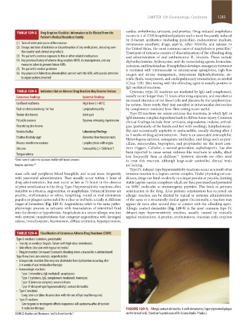
physiologic process as urticaria with transudation of interstitial fluid Allergic contact dermatitis (Fig. 129-5) is the most common type IV,
into the dermis or hypodermis. Anaphylaxis is a severe allergic reaction delayed-type hypersensitivity reaction, usually caused by topically
with systemic manifestations that comprise angioedema with laryngeal applied medications. A pruritic, erythematous, vesicular, scaly eruption
edema, bronchospasm, hypotension, diffuse erythema, hyperperistalsis,
TABLE 129-4 Classification of Cutaneous Adverse Drug Reactions (CADR)
Type A reactions (common, predictable)
• Toxicity or overdose (hepatic failure with high-dose isotretinoin)
• Side effects (dry skin with topical retinoids)
• Drug interaction (increased Coumadin bleeding when a macrolide is administered)
Type B reactions (uncommon, unpredictable)
• Idiosyncratic reaction (the very rare cholestatic liver dysfunction occurring after
3-4 weeks of oral terbinafine therapy)
• Immunologic reactions
Type I (immediate, IgE mediated): anaphylaxis
Type II (cytotoxic, IgG, complement-mediated): hemolysis
Type III (immune complex): serum sickness
Type IV (delayed-type hypersensitivity): contact dermatitis
• Type C reactions
Long-term use (blue discoloration with the use of hydroxychloroquine)
• Type D reactions
Carcinogenic or teratogenic effects (squamous cell carcinoma after ultraviolet
A radiation therapy) FIGURE 129-5. Allergic contact dermatitis. A well-demarcated, hyperpigmented plaque
SOURCES: Rawlins and Thompson, and Gell and Coombs. 8 on the lateral neck. (Used with permission of Dr Juliana Basko-Plluska.)
5
section11.indd 1283 1/19/2015 10:52:42 AM