
Page 1811 - Hall et al (2015) Principles of Critical Care-McGraw-Hill
P. 1811
1280 PART 11: Special Problems in Critical Care
CHAPTER Dermatologic Conditions layers of the skin. Preparing the skin for invasive procedures with topi-
cal solutions exposes the patient to potential sensitizers. Metabolically
129 Juliana L. Basko-Plluska active cells in the suprabasal layers are susceptible to inflammatory
and cytotoxic reactions from medications and toxins. Disruption
Rekha Vij
Aisha Sethi in cell adhesion clinically manifests as blisters and may result from
medications, toxins, pressure, extremes in temperature, or autoimmune
diseases. Infections and inflammatory processes can occur at any level
or in any structure, leading to conditions such as impetigo, folliculitis,
KEY POINTS
cellulitis, fasciitis, or vasculitis.
• In a patient with a dermatologic condition, observation and ■
description of the lesions (morphology, distribution, and texture) BASIC MORPHOLOGIC APPROACH AND DESCRIPTIONS
are important for developing a differential diagnosis. When approaching a patient with skin disease, careful observation,
• Mucous membranes (oral, ocular, nasal, genital, and perianal) palpation, and description are critical for developing a differential diag-
should be examined in all patients. nosis. The morphology, or type, of lesion may be flat (macule), elevated
• The skin may provide clues to an underlying, life-threatening (papule, nodule, plaque, cyst, vesicle, bulla, pustule, or hyperkeratosis),
condition, such as endocarditis, graft-versus-host disease, bacterial or depressed (ulcer or atrophy; Table 129-1). Shape, margination
and fungal sepsis, toxic shock syndrome, systemic vasculitis, or (well or poorly defined borders), and arrangement of the lesions are
complications from the human immunodeficiency virus. important. Color may be white (leukoderma or hypomelanosis), red
• Drug-related dermatoses are prevalent in the intensive care unit. (erythema), pink, violaceous (purple), brown (hypermelanosis or hemo-
siderin), black, blue, gray, or yellow. Particular attention should be paid
Clues to diagnosis include a rapidly developing eruption; general- to the distribution of the eruption (eg, localized, systemic, truncal, acral,
ized, symmetrical, predominantly truncal distribution; morbilliform, unilateral, or intertriginous). Palpation will help determine consistency,
urticarial, or acneiform morphology; and accompanying pruritus. temperature, mobility, tenderness, and depth of lesion. When various
• Extensive skin disease can cause important fluid, electrolyte, and pro- lesions are present, one should attempt to determine the primary lesion.
tein losses and predisposes the patient to life-threatening infections. Clinical history is essential. Cutaneous symptoms (pruritus, pain,
tenderness, burning, or stinging) as well as systemic symptoms (fever,
malaise, arthralgias, myalgias, etc) must be ascertained. Time course
of the skin lesions should be determined, with particular attention to
BASICS OF DERMATOLOGY medication history (prescription and nonprescription, oral, topical, and
■ APPLICATION OF STRUCTURE AND FUNCTION TO DERMATOSES alternative). Common diagnostic aids in dermatology are skin biopsy
for hematoxylin and eosin or other special staining, direct immuno-
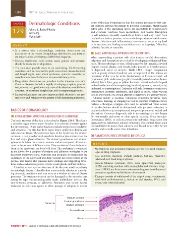
The basic anatomy of the skin is described in Figure 129-1. The skin is fluorescence (DIF), or culture; potassium hydroxide preparation (for
a complex organ whose major function is to provide a barrier against dermatophytic infections); mineral oil mounts (for scabies); Gram stain
the environment. Other major functions include temperature regulation (for bacterial infections); fluid cultures; and Tzanck smears (for herpes
and sensation. The skin has three major layers: epidermis, dermis, and simplex and varicella zoster virus infections).
subcutaneous tissue. The outermost layer of the epidermis, the stratum
corneum, is composed of dead, anucleate keratinocytes and serves as the DERMATOSES PRECIPITATED BY DRUGS
first and major physical barrier. The stratum granulosum and stratum
spinosum lie below the stratum corneum and are composed of keratino- KEY POINTS
cytes in the process of differentiation. They are derived from the bottom
layer of the epidermis, the basal cell layer. The epidermis is connected • Morbilliform and urticarial eruptions are the two most common
to the dermis by a complex of proteins and adhesion molecules in the types of drug eruptions.
basement membrane zone. Nutrients and products of metabolism are • Less common reactions include pustular, bullous, vasculitic,
exchanged in the superficial and deep vascular networks located in the lichenoid, and fixed drug eruptions.
dermis. The dermis also contains nerve endings and supporting struc-
tures such as sebaceous glands, eccrine sweat glands, and hair follicles. • Stevens-Johnson syndrome (SJS), toxic epidermal necrolysis
Alteration of any layer or structure of the skin can result in primary (TEN), and drug reaction with eosinophilia and systemic symp-
dermatologic disease. Often the skin is secondarily affected in underly- toms (DRESS) are three severe cutaneous drug reactions that need
ing comorbid conditions and may serve as a window to internal disease prompt recognition and initiation of treatment.
processes. The stratum corneum can be damaged in the intensive care • Therapy consists of withdrawal of the culprit drug, symptomatic
setting by tape, electrocardiographic leads, defibrillator devices, dry relief with antihistamines ± topical or oral steroids, IVIG, and
environments, pressure, or adhesives. Alteration may impair barrier wound care when indicated.
resistance to infectious agents or allow passage of antigens to deeper
Stratum corneum
Stratum granulosum
Epidermis
Stratum spinosum
Stratum basale
Dermis
FIGURE 129-1. Structure of normal skin. (Used with permission of Dr Jie Song.)
section11.indd 1280 1/19/2015 10:52:31 AM