
Page 1816 - Hall et al (2015) Principles of Critical Care-McGraw-Hill
P. 1816
CHAPTER 129: Dermatologic Conditions 1285
and topotecan have also been implicated. NEH typically resolves within
TABLE 129-6 Antidotes Used in Chemotherapeutic Agent Extravasation
weeks after cessation of chemotherapy, although it may recur in subse-
Agent Antidote quent cycles. Dapsone is used to treat and prevent recurrences of NEH. 22
Anthracycline Topical DMSO ■
Mitomycin Topical DMSO ANTICOAGULANT-INDUCED DERMATOSES
Adverse reactions to anticoagulants can be localized or diffuse and
Vinca alkaloid Hyaluronidase (ICA)
mediated by various mechanisms. Unfractionated heparin, low-molecu-
Mechlorethamine Sodium thiosulfate (ICA) lar-weight heparins, and vitamin K antagonists are drugs used routinely
Dacarbazine Sodium thiosulfate (ICA) in the treatment and prophylaxis of thromboembolic disease. In addi-
Cisplatin Sodium thiosulfate (ICA) tion to the well-known ADRs such as an increased bleeding tendency,
these agents may cause CADR which necessitate institution of alterna-
DMSO, dimethylsulfoxide; ICA, intracutaneous administration.
tive therapies.
Data from Susser WS, Whitaker-Worth DL, Grant-Kels JM. Mucocutaneous reactions to chemotherapy. Heparin is a mucopolysaccharide that can induce various ADRs,
J Am Acad Dermatol. March 1999;40(3):367-398. including immediate reactions (eg, urticaria, asthma, and anaphylaxis),
and delayed-type reactions (eg, erythematous plaques, skin necrosis, a
23
with bleomycin, cytarabine, cyclophosphamide, daunorubicin, doxo- generalized eruption, and thrombocytopenia). Delayed-type reactions
rubicin, fluorouracil, hydroxyurea, idarubicin, mitomycin, tegafur, or manifest as erythematous, vesicular, or pruritic plaques at the injection
vinblastine; and mucosal hyperpigmentation with busulfan, fluorouracil, site within 2 to 5 days of therapy, whereas heparin-induced skin necrosis
tegafur, doxorubicin, hydroxyurea, cisplatin, or cyclophosphamide. 13 (Fig. 129-7) develops between 5 and 10 days of therapy. Cutaneous reac-
Another well-described CADR to chemotherapeutic agents is acral tions have also been described with the use of low-molecular-weight
24
erythema (Fig. 129-6), occurring in 6% to 24% of patients, mainly heparins and heparinoids. In patients with skin necrosis, heparin must
18
those treated with cytarabine, doxorubicin, and fluorouracil. This pain- be discontinued due to the increased risk of developing heparin-induced
25
ful eruption consists of diffuse erythema and edema on the palms and thrombocytopenia II. These patients usually have significant concurrent
soles. This resolves with exfoliation within 4 weeks of discontinuing the illnesses but no coagulation abnormalities. Histology shows fibrin deposi-
drug. It needs to be differentiated from acute GVHD in the appropriate tion in venules and capillaries in the dermis and hypodermis, with necrosis.
clinical setting. Treatment is supportive, with elevation, cold compresses, Intracutaneous testing is useful for the diagnosis of delayed reactions
analgesics, and pyridoxine 150 mg each day. 19 (erythematous plaques at heparin injection sites) but is contraindicated
Cutaneous ulceration of the lower extremities is reported as a compli- in the presence of skin necrosis. A positive skin test has been associated
cation of long-term hydroxyurea, and rarely, high-dose methotrexate. with heparin-induced IgG antibodies. There may be cross-reactivity
20
21
Hydroxyurea-induced ulcers typically occur over the malleoli, are between unfractionated heparin and low-molecular-weight heparins and
painful, and may be resistant to local wound care, topical or systemic heparinoids. Rates of 50% and 30%, respectively, have been reported.
26
antibiotic therapy, pentoxifylline, prednisone, hyperbaric oxygen, or Therefore, treatment consists of discontinuing heparin and administering
Unna vascular boots. Ulcers heal over several months after cessation of one of the newly developed direct thrombin inhibitors (argatroban and
hydroxyurea. The first case of lower extremity cutaneous ulceration in a lepirudin) to provide protection during warfarin initiation.
patient without psoriasis receiving methotrexate was reported in 1998. Warfarin inhibits the vitamin K–dependent clotting factors II, VII, IX,
21
Since then, several additional cases of methotrexate-related cutaneous and X; it also inhibits the anticoagulant proteins C and S, thereby caus-
ulcers have been reported. ing a transient hypercoagulable state. On rare occasions, skin necrosis
27
Neutrophilic eccrine hidradenitis (NEH) is a disorder that results (Fig. 129-8), which is potentially fatal (15% of cases), occurs with initia-
from a direct cytotoxic effect of chemotherapy on the eccrine glands. tion of therapy before full anticoagulation is achieved. Three to ten days
Erythematous papules and plaques occur on the trunk and the extremi- after starting therapy, pain precedes the appearance of ill-defined ery-
ties within 2 weeks of starting chemotherapy. This condition is most often thematous plaques, which progress into edematous, blue-black plaques
associated with the initiation of cytarabine in patients with acute myelog- with hemorrhagic bullae in the center. The lesions necrose, leaving central
enous leukemia. Bleomycin, cyclophosphamide, anthracyclines, cisplatin, eschars. Necrosis commonly affects the breasts, abdomen, and thighs in
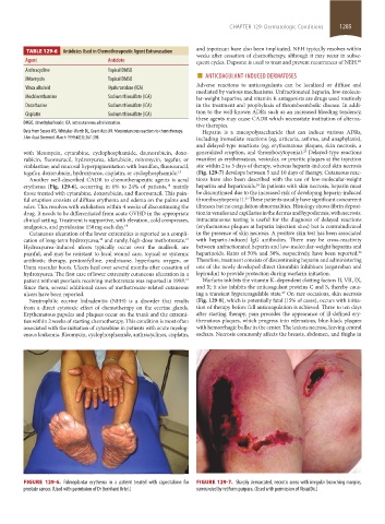
FIGURE 129-6. Palmoplantar erythema in a patient treated with capecitabine for FIGURE 129-7. Sharply demarcated, necrotic areas with irregular branching margins,
prostate cancer. (Used with permission of Dr Bernhard Ortel.) surrounded by retiform purpura. (Used with permission of VisualDx.)
section11.indd 1285 1/19/2015 10:52:51 AM