


Page 122 - Textbook of Pathology, 6th Edition
P. 122
106


     SECTION I


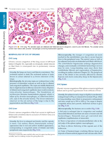
           Figure 5.10  CVC lung. The alveolar septa are widened and thickened due to congestion, oedema and mild fibrosis. The alveolar lumina
           contain heart failure cells (alveolar macrophages containing haemosiderin pigment).

           MORPHOLOGY OF CVC OF ORGANS                           Microscopically, the changes of congestion are more
                                                                 marked in the centrilobular zone due to severe hypoxia
           CVC Lung                                              than in the peripheral zone. The central veins as well as
           Chronic venous congestion of the lung occurs in left heart  the adjacent sinusoids are distended and filled with blood.
           failure (Chapter 16), especially in rheumatic mitral stenosis  The centrilobular hepatocytes undergo degenerative
           so that there is consequent rise in pulmonary venous  changes, and eventually centrilobular haemorrhagic necrosis
           pressure.                                             may be seen. Long-standing cases may show fine
                                                                 centrilobular fibrosis and regeneration of hepatocytes,
            Grossly, the lungs are heavy and firm in consistency. The  resulting in cardiac cirrhosis (Chapter 21). The peripheral
     General Pathology and Basic Techniques
            sectioned surface is dark The sectioned surface is rusty  zone of the lobule is less severely affected by chronic
            brown in colour referred to as  brown induration of the  hypoxia and shows some fatty change in the hepatocytes
            lungs.                                               (Fig. 5.12).
            Histologically, the alveolar septa are widened due to the
            presence of interstitial oedema as well as due to dilated
            and congested capillaries. The septa are mildly thickened  CVC Spleen
            due to slight increase in fibrous connective tissue. Rupture  Chronic venous congestion of the spleen occurs in right heart
            of dilated and congested capillaries may result in minute  failure and in portal hypertension from cirrhosis of liver.
            intra-alveolar haemorrhages. The breakdown of
            erythrocytes liberates haemosiderin pigment which is  Grossly, the spleen in early stage is slightly to moderately
            taken up by alveolar macrophages, so called heart failure  enlarged (up to 250 g as compared to normal 150 g), while
            cells, seen in the alveolar lumina. The brown induration  in long-standing cases there is progressive enlargement
            observed on the cut surface of the lungs is due to the  and may weigh up to 500 to 1000 g. The organ is deeply
            pigmentation and fibrosis (Fig. 5.10).               congested, tense and cyanotic. Sectioned surface is gray
                                                                 tan (Fig. 5.13).
           CVC Liver                                             Microscopically, the features are as under (Fig. 5.14):
                                                                 i) Red pulp is enlarged due to congestion and marked
           Chronic venous congestion of the liver occurs in right heart
           failure and sometimes due to occlusion of inferior vena cava  sinusoidal dilatation and here are areas of recent and old
           and hepatic vein.                                     haemorrhages. Sinusoids may get converted into
                                                                 capillaries (capillarisation of sinusoids).
            Grossly, the liver is enlarged and tender and the capsule  ii) There is hyperplasia of reticuloendothelial cells in the
            is tense. Cut surface shows characteristic  nutmeg*  red pulp of the spleen (splenic macrophages).
            appearance due to red and yellow mottled appearance,  iii) There is fibrous thickening of the capsule and of the
            corresponding to congested centre of lobules and fatty  trabeculae.
            peripheral zone respectively (Fig. 5.11).
                                                                 iv) Some of haemorrhages overlying fibrous tissue get
                                                                 deposits of haemosiderin pigment and calcium salts; these
           *Nutmeg (vernacular name jaiphal) is the seed of a spice tree that grows
           in India, used in cooking as spice for giving flavours.

