


Page 121 - Textbook of Pathology, 6th Edition
P. 121
DISTURBANCES IN THE VOLUME OF                       Passive Hyperaemia (Venous Congestion)               105
           CIRCULATING BLOOD
                                                               The dilatation of veins and capillaries due to impaired venous
           HYPERAEMIA AND CONGESTION                           drainage results in passive hyperaemia or venous congestion,
           Hyperaemia and congestion are the terms used for localised  commonly referred to as congestion. Congestion may be acute
           increase in the volume of blood within dilated vessels of an  or chronic, the latter being more common and called chronic
           organ or tissue; the increased volume from arterial and  venous congestion (CVC). The affected tissue or organ is bluish  CHAPTER 5
           arteriolar dilatation being referred to as hyperaemia or active  in colour due to accumulation of venous blood (cyanosis).
           hyperaemia, whereas the impaired venous drainage is called  Obstruction to the venous outflow may be local or systemic.
           venous congestion or passive hyperaemia. If the condition  Accordingly, venous congestion is of 2 types:
           develops rapidly it is called acute, while more prolonged and  Local venous congestion results from obstruction to the
           gradual response is known as chronic.               venous outflow from an organ or part of the body e.g. portal
           Active Hyperaemia                                   venous obstruction in cirrhosis of the liver, outside pressure
           The dilatation of arteries, arterioles and capillaries is effected  on the vessel wall as occurs in tight bandage, plasters,
                                                               tumours, pregnancy, hernia etc, or intraluminal occlusion
           either through sympathetic neurogenic mechanism or via the  by thrombosis.
           release of vasoactive substances. The affected tissue or organ
           is pink or red in appearance (erythema).                Systemic (General) venous congestion is engorgement of
              The  examples of active hyperaemia are seen in the  systemic veins e.g. in left-sided and right-sided heart failure
           following conditions:                               and diseases of the lungs which interfere with pulmonary
              Inflammation e.g. congested vessels in the walls of alveoli  blood flow like pulmonary fibrosis, emphysema etc. Usually
           in pneumonia                                        the fluid accumulates upstream to the specific chamber of
              Blushing i.e. flushing of the skin of face in response to  the heart which is initially affected (Chapter 16). For
           emotions                                            example, in left-sided heart failure (such as due to mechanical
              Menopausal flush                                 overload in aortic stenosis, or due to weakened left   Derangements of Homeostasis and Haemodynamics
              Muscular exercise                                ventricular wall as in myocardial infarction) pulmonary
              High grade fever                                 congestion results, whereas in right-sided heart failure (such
              Goitre                                           as due to pulmonary stenosis or pulmonary hypertension)
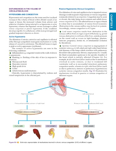
              Arteriovenous malformations                      systemic venous congestion results. Fig. 5.9 illustrates the
              Clinically, hyperaemia is characterised by redness and  mechanisms involved in passive or venous congestion of
           raised temperature in the affected part.            different organs.


           Figure 5.9  Schematic representation of mechanisms involved in chronic venous congestion (CVC) of different organs.

