


Page 123 - Textbook of Pathology, 6th Edition
P. 123
107


                                                                                                                      CHAPTER 5


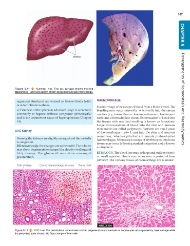
           Figure 5.11  Nutmeg liver.  The cut surface shows mottled
           appearance—alternate pattern of dark congestion and pale fatty change.


                                                               HAEMORRHAGE                                            Derangements of Homeostasis and Haemodynamics
            organised structures are termed as Gamna-Gandy bodies
            or siderofibrotic nodules.
                                                               Haemorrhage is the escape of blood from a blood vessel. The
            v) Firmness of the spleen in advanced stage is seen more  bleeding may occur externally, or internally into the serous
            commonly in hepatic cirrhosis (congestive splenomegaly)  cavities (e.g. haemothorax, haemoperitoneum, haemoperi-
            and is the commonest cause of hypersplenism (Chapter  cardium), or into a hollow viscus. Extravasation of blood into
            14).                                               the tissues with resultant swelling is known as haematoma.
                                                               Large extravasations of blood into the skin and mucous
                                                               membranes are called ecchymoses. Purpuras are small areas
           CVC Kidney
                                                               of haemorrhages (upto 1 cm) into the skin and mucous
                                                               membrane, whereas  petechiae are minute pinhead-sized
            Grossly, the kidneys are slightly enlarged and the medulla  haemorrhages. Microscopic escape of erythrocytes into loose
            is congested.                                      tissues may occur following marked congestion and is known
            Microscopically, the changes are rather mild. The tubules  as diapedesis.
            may show degenerative changes like cloudy swelling and
            fatty change. The glomeruli may show mesangial     ETIOLOGY. The blood loss may be large and sudden (acute),
            proliferation.                                     or small repeated bleeds may occur over a period of time
                                                               (chronic). The various causes of haemorrhage are as under:


           Figure 5.12  CVC liver. The centrilobular zone shows marked degeneration and necrosis of hepatocytes accompanied by haemorrhage while
           the peripheral zone shows mild fatty change of liver cells.

