


Page 151 - Textbook of Pathology, 6th Edition
P. 151
135


                                                                                                                      CHAPTER 6


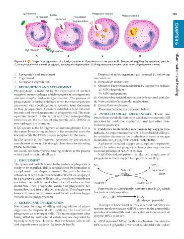
           Figure 6.6  Stages in phagocytosis of a foreign particle. A, Opsonisation of the particle. B, Pseudopod engulfing the opsonised particle.
           C, Incorporation within the cell (phagocytic vacuole) and degranulation. D, Phagolysosome formation after fusion of lysosome of the cell.


           1. Recognition and attachment                          Disposal of microorganisms can proceed by following  Inflammation and Healing
           2. Engulfment                                       mechanisms:
           3. Killing and degradation                          A. Intracellular mechanisms:
                                                               i) Oxidative bactericidal mechanism by oxygen free radicals
           1. RECOGNITION AND ATTACHMENT
           Phagocytosis is initiated by the expression of surface  a) MPO-dependent
           receptors on macrophages which recognise microorganisms:  b) MPO-independent
           mannose receptor and  scavenger receptor. The process of  ii) Oxidative bactericidal mechanism by lysosomal granules
           phagocytosis is further enhanced when the microorganisms  iii) Non-oxidative bactericidal mechanism
           are coated with specific proteins, opsonins, from the serum  B. Extracellular mechanisms:
           or they get opsonised. Opsonins establish a bond between  These mechanisms are discussed below.
           bacteria and the cell membrane of phagocytic cell. The main  A. INTRACELLULAR MECHANISMS. There are
           opsonins present in the serum and their corresponding  intracellular metabolic pathways which more commonly kill
           receptors on the surface of phagocytic cells (PMNs or  microbes by oxidative mechanism and less often non-
           macrophages) are as under:
                                                               oxidative pathways.
           i) IgG opsonin is the Fc fragment of immunoglobulin G; it is  i) Oxidative bactericidal mechanism by oxygen free
           the naturally occurring antibody in the serum that coats the  radicals. An important mechanism of microbicidal killing is
           bacteria while the PMNs possess receptors for the same.  by oxidative damage by the production of reactive oxygen
           ii) C3b opsonin is the fragment generated by activation of  metabolites (O’  H O , OH’, HOCl, HOI, HOBr).
                                                                                 2
                                                                            2
                                                                               2
           complement pathway. It is strongly chemotactic for attracting  A phase of increased oxygen consumption (‘respiratory
           PMNs to bacteria.                                   burst’) by activated phagocytic leucocytes requires the
           iii) Lectins are carbohydrate-binding proteins in the plasma  essential presence of NADPH oxidase.
           which bind to bacterial cell wall.                     NADPH-oxidase present in the cell membrane of
                                                               phagosome reduces oxygen to superoxide ion (O’ ):
           2. ENGULFMENT                                                                                  2
           The opsonised particle bound to the surface of phagocyte is  2O                            2O’
           ready to be engulfed. This is accomplished by formation of  2                                 2
           cytoplasmic pseudopods around the particle due to                     NADPH               (Superoxide
           activation of actin filaments beneath cell wall, enveloping it        oxidase                   anion)
           in a phagocytic vacuole. Eventually, the plasma membrane
           enclosing the particle breaks from the cell surface so that   NADPH                        NADP + H +
           membrane lined phagocytic vacuole or phagosome lies
           internalised and free in the cell cytoplasm. The phagosome  Superoxide is subsequently converted into H O  which
                                                                                                           2
                                                                                                         2
           fuses with one or more lysosomes of the cell and form bigger  has bactericidal properties:
           vacuole called phagolysosome.                             2O’  + 2H +            H O 2
                                                                                              2
                                                                        2
                                                                                            (Hydrogen peroxide)
           3. KILLING AND DEGRADATION
           Next comes the stage of killing and degradation of micro-  This type of bactericidal activity is carried out either via
           organism to dispose it off justifying the function of  enzyme myeloperoxidase (MPO) present in the azurophilic
           phagocytes as scavanger cells. The microorganisms after  granules of neutrophils and monocytes, or independent of
           being killed by antibacterial substances are degraded by  enzyme MPO, as under:
           hydrolytic enzymes. However, this mechanism fails to kill  a) MPO-dependent killing. In this mechanism, the enzyme
           and degrade some bacteria like tubercle bacilli.    MPO acts on H O  in the presence of halides (chloride, iodide
                                                                            2
                                                                              2

