


Page 150 - Textbook of Pathology, 6th Edition
P. 150
134 stasis, the central stream of cells widens and peripheral
           plasma zone becomes narrower because of loss of plasma
           by exudation. This phenomenon is known as margination.
           As a result of this redistribution, the neutrophils of the central
           column come close to the vessel wall; this is known as
           pavementing.
           2. ROLLING AND ADHESION. Peripherally marginated
           and pavemented neutrophils slowly roll over the endothelial
           cells lining the vessel wall (rolling phase).  This is followed by
     SECTION I
           the transient bond between the leucocytes and endothelial
           cells becoming firmer  (adhesion phase).  The following
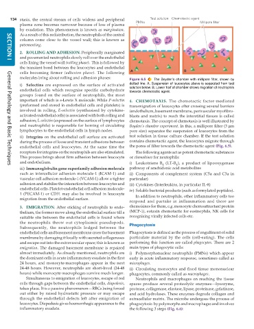
           molecules bring about rolling and adhesion phases:  Figure 6.5  The Boyden’s chamber with millipore filter, shown by
           i) Selectins  are expressed on the surface of activated  dotted line. A, Suspension of leucocytes above is separated from test
           endothelial cells which recognise specific carbohydrate  solution below. B, Lower half of chamber shows migration of neutrophils
                                                               towards chemotactic agent.
           groups found on the surface of neutrophils, the most
           important of which is s-Lewis X molecule. While P-selectin  4. CHEMOTAXIS.  The chemotactic factor-mediated
           (preformed and stored in endothelial cells and platelets) is  transmigration of leucocytes after crossing several barriers
           involved in rolling,  E-selectin (synthesised by cytokine-  (endothelium, basement membrane, perivascular myofibro-
           activated endothelial cells) is associated with both rolling and  blasts and matrix) to reach the interstitial tissues is called
           adhesion; L-selectin (expressed on the surface of lymphocytes  chemotaxis. The concept of chemotaxis is well illustrated by
           and neutrophils) is responsible for homing of circulating  Boyden’s chamber experiment. In this, a millipore filter (3 μm
           lymphocytes to the endothelial cells in lymph nodes.  pore size) separates the suspension of leucocytes from the
           ii) Integrins on the endothelial cell surface are activated  test solution in tissue culture chamber. If the test solution
           during the process of loose and transient adhesions between  contains chemotactic agent, the leucocytes migrate through
           endothelial cells and leucocytes. At the same time the  the pores of filter towards the chemotactic agent (Fig. 6.5).
           receptors for integrins on the neutrophils are also stimulated.  The following agents act as potent chemotactic substances
           This process brings about firm adhesion between leucocyte  or chemokines for neutophils:
           and endothelium.                                    i) Leukotriene B  (LT-B ), a product of lipooxygenase
                                                                               4
                                                                                      4
     General Pathology and Basic Techniques
           iii) Immunoglobulin gene superfamily adhesion molecule  pathway of arachidonic acid metabolites
           such as intercellular adhesion molecule-1 (ICAM-1) and  ii) Components of complement system (C5a and C3a in
           vascular cell adhesion molecule-1 (VCAM-1) allow a tighter  particular)
           adhesion and stabilise the interaction between leucocytes and  iii) Cytokines (Interleukins, in particular IL-8)
           endothelial cells. Platelet-endothelial cell adhesion molecule-  iv) Soluble bacterial products (such as formylated peptides).
           1 (PECAM-1) or CD31 may also be involved in leucocyte  In addition to neutrophils, other inflammatory cells too
           migration from the endothelial surface.
                                                               respond and partake in inflammation and there are
           3. EMIGRATION. After sticking of neutrophils to endo-  chemokines for them, e.g. monocyte chemoattractant protein
           thelium, the former move along the endothelial surface till a  (MCP-1), eotaxin chemotactic for eosinophils, NK cells for
           suitable site between the endothelial cells is found where  recognising virally infected cells etc.
           the neutrophils throw out cytoplasmic pseudopods.   Phagocytosis
           Subsequently, the neutrophils lodged between the
           endothelial cells and basement membrane cross the basement  Phagocytosis is defined as the process of engulfment of solid
           membrane by damaging it locally with secreted collagenases  particulate material by the cells (cell-eating). The cells
           and escape out into the extravascular space; this is known as  performing this function are called phagocytes. There are 2
           emigration. The damaged basement membrane is repaired  main types of phagocytic cells:
           almost immediately. As already mentioned, neutrophils are  i) Polymorphonuclear neutrophils (PMNs) which appear
           the dominant cells in acute inflammatory exudate in the first  early in acute inflammatory response, sometimes called as
           24 hours, and monocyte-macrophages appear in the next  microphages.
           24-48 hours. However, neutrophils are short-lived (24-48  ii) Circulating monocytes and fixed tissue mononuclear
           hours) while monocyte-macrophages survive much longer.  phagocytes, commonly called as macrophages.
              Simultaneous to emigration of leucocytes, escape of red  Neutrophils and macrophages on reaching the tissue
           cells through gaps between the endothelial cells, diapedesis,  spaces produce several proteolyitc enzymes—lysozyme,
           takes place. It is a passive phenomenon—RBCs being forced  protease, collagenase, elastase, lipase, proteinase, gelatinase,
           out either by raised hydrostatic pressure or may escape  and acid hydrolases. These enzymes degrade collagen and
           through the endothelial defects left after emigration of  extracellular matrix. The microbe undergoes the process of
           leucocytes. Diapedesis gives haemorrhagic appearance to the  phagocytosis  by polymorphs and macrophages and involves
           inflammatory exudate.                               the following 3 steps (Fig. 6.6):

