


Page 156 - Textbook of Pathology, 6th Edition
P. 156
140


     SECTION I


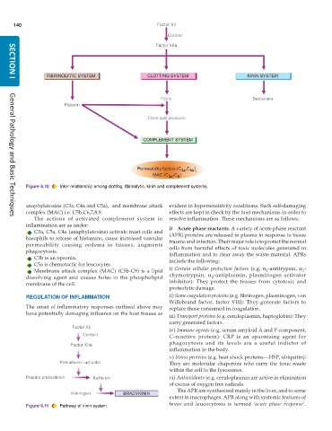
           Figure 6.10  Inter-relationship among clotting, fibrinolytic, kinin and complement systems.


           anaphylatoxins (C3a, C4a and C5a),  and membrane attack  evident in hypersensitivity conditions. Such self-damaging


           complex (MAC) i.e. C5b,C6,7,8,9.                    effects are kept in check by the host mechanisms in order to
              The actions of activated complement system in    resolve inflammation. These mechanisms are as follows:
     General Pathology and Basic Techniques
           inflammation are as under:                          i) Acute phase reactants. A variety of acute phase reactant
              C3a, C5a, C4a (anaphylatoxins) activate mast cells and
           basophils to release of histamine, cause increased vascular  (APR) proteins are released in plasma in response to tissue
                                                               trauma and infection. Their major role is to protect the normal
           permeability causing oedema in tissues, augments    cells from harmful effects of toxic molecules generated in
           phagocytosis.                                       inflammation and to clear away the waste material. APRs
              C3b is an opsonin.                               include the following:
              C5a is chemotactic for leucocytes.
              Membrane attack complex (MAC) (C5b-C9) is a lipid  i) Certain cellular protection factors (e.g.  α -antitrypsin,  α -
                                                                                                   1
                                                                                                                1
           dissolving agent and causes holes in the phospholipid  chymotrypsin,  α -antiplasmin, plasminogen activator
                                                                               2
           membrane of the cell.                               inhibitor): They protect the tissues from cytotoxic and
                                                               proteolytic damage.
           REGULATION OF INFLAMMATION                          ii) Some coagulation proteins (e.g. fibrinogen, plasminogen, von
                                                               Willebrand factor, factor VIII): They generate factors to
           The onset of inflammatory responses outlined above may  replace those consumed in coagulation.
           have potentially damaging influence on the host tissues as
                                                               iii) Transport proteins (e.g. ceruloplasmin, haptoglobin): They
                                                               carry generated factors.
                                                               iv) Immune agents (e.g. serum amyloid A and P component,
                                                               C-reactive protein): CRP is an opsonising agent for
                                                               phagocytosis and its levels are a useful indictor of
                                                               inflammation in the body.
                                                               v) Stress proteins (e.g. heat shock proteins—HSP, ubiquitin):
                                                               They are molecular chaperons who carry the toxic waste
                                                               within the cell to the lysosomes.
                                                               vi) Antioxidants (e.g. ceruloplasmin are active in elimination
                                                               of excess of oxygen free radicals.
                                                                  The APR are synthesised mainly in the liver, and to some
                                                               extent in macrophages. APR along with systemic features of

           Figure 6.11  Pathway of kinin system.               fever and leucocytosis is termed  ‘acute phase response’.

