


Page 158 - Textbook of Pathology, 6th Edition
P. 158
142
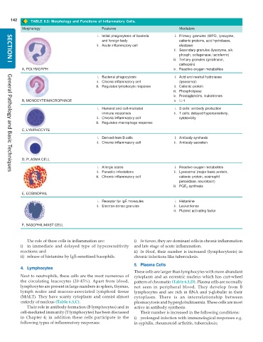
TABLE 6.3: Morphology and Functions of Inflammatory Cells.
Morphology Features Mediators
i. Initial phagocytosis of bacteria i. Primary granules (MPO, lysozyme,
and foreign body cationic proteins, acid hydrolases,
ii. Acute inflammatory cell elastase)
ii. Secondary granules (lysozyme, alk.
phosph, collagenase, lactoferrin)
iii. Tertiary granules (gelatinase,
cathepsin)
SECTION I
A, POLYMORPH iv. Reactive oxygen metabolites
i. Bacterial phagocytosis i. Acid and neutral hydrolases
ii. Chronic inflammatory cell (lysosomal)
iii. Regulates lymphocyte response ii. Cationic protein
iii. Phospholipase
iv. Prostaglandins, leukotrienes
B, MONOCYTE/MACROPHAGE v. IL-1
i. Humoral and cell-mediated i. B cells: antibody production
immune responses ii. T cells: delayed hypersensitivity,
ii. Chronic inflammatory cell cytotoxicity
iii. Regulates macrophage response
C, LYMPHOCYTE
i. Derived from B cells i. Antibody synthesis
ii. Chronic inflammatory cell ii. Antibody secretion
D, PLASMA CELL
i. Allergic states i. Reactive oxygen metabolites
ii. Parasitic infestations ii. Lysosomal (major basic protein,
General Pathology and Basic Techniques
iii. Chronic inflammatory cell cationic protein, eosinophil
peroxidase, neurotoxin)
iii. PGE 2 synthesis
E, EOSINOPHIL
i. Receptor for IgE molecules i. Histamine
ii. Electron-dense granules ii. Leukotrienes
iii. Platelet activating factor
F, BASOPHIL/MAST CELL
The role of these cells in inflammation are: i) In tissues, they are dominant cells in chronic inflammation
i) in immediate and delayed type of hypersensitivity and late stage of acute inflammation.
reactions; and ii) In blood, their number is increased (lymphocytosis) in
ii) release of histamine by IgE-sensitised basophils. chronic infections like tuberculosis.
5. Plasma Cells
4. Lymphocytes
These cells are larger than lymphocytes with more abundant
Next to neutrophils, these cells are the most numerous of cytoplasm and an eccentric nucleus which has cart-wheel
the circulating leucocytes (20-45%). Apart from blood, pattern of chromatin (Table 6.3,D). Plasma cells are normally
lymphocytes are present in large numbers in spleen, thymus, not seen in peripheral blood. They develop from B
lymph nodes and mucosa-associated lymphoid tissue lymphocytes and are rich in RNA and γ-globulin in their
(MALT). They have scanty cytoplasm and consist almost cytoplasm. There is an interrelationship between
entirely of nucleus (Table 6.3,C). plasmacytosis and hyperglobulinaemia. These cells are most
Their role in antibody formation (B lymphocytes) and in active in antibody synthesis.
cell-mediated immunity (T lymphocytes) has been discussed Their number is increased in the following conditions:
in Chapter 4; in addition these cells participate in the i) prolonged infection with immunological responses e.g.
following types of inflammatory responses: in syphilis, rheumatoid arthritis, tuberculosis;

