


Page 159 - Textbook of Pathology, 6th Edition
P. 159
143


                                                                                                                      CHAPTER 6


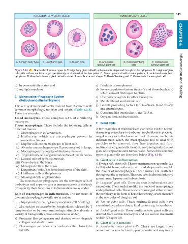
           Figure 6.14  Giant cells of various types. A, Foreign body giant cell with uniform nuclei dispersed throughout the cytoplasm. B, Langhans’ giant
           cells with uniform nuclei arranged peripherally or clustered at the two poles. C, Touton giant cell with circular pattern of nuclei and vacuolated
           cytoplasm. D, Anaplastic tumour giant cell with nuclei of variable size and shape. E, Reed-Sternberg cell. F, Osteoclastic tumour giant cell.  Inflammation and Healing


           ii) hypersensitivity states; and                    c) Products of complement.
           iii) multiple myeloma.                              d) Some coagulation factors (factor V and thromboplastin)
                                                                  which convert fibrinogen to fibrin.
           6. Mononuclear-Phagocyte System                     e) Chemotactic agents for other leucocytes.
              (Reticuloendothelial System)                     f) Metabolites of arachidonic acid.
           This cell system includes cells derived from 2 sources with  g) Growth promoting factors for fibroblasts, blood vessels
           common morphology, function and origin  (Table 6.3,B).  and granulocytes.
           These are as under:                                 h) Cytokines like interleukin-1 and TNF-α.
           Blood monocytes. These comprise 4-8% of circulating  i) Oxygen-derived free radicals.
           leucocytes.                                         7. Giant Cells
           Tissue macrophages. These include the following cells in
           different tissues:                                  A few examples of multinucleate giant cells exist in normal
            i) Macrophages in inflammation.                    tissues (e.g. osteoclasts in the bones, trophoblasts in placenta,
            ii) Histiocytes which are macrophages present in   megakaryocytes in the bone marrow). However, in chronic
               connective tissues.                             inflammation when the macrophages fail to deal with
           iii) Kupffer cells are macrophages of liver cells.  particles to be removed, they fuse together and form
           iv) Alveolar macrophages (type II pneumocytes) in lungs.  multinucleated giant cells. Besides, morphologically distinct
            v) Macrophages/histiocytes of the bone marrow.     giant cells appear in some tumours also. Some of the common
           vi) Tingible body cells of germinal centres of lymph nodes.  types of giant cells are described below (Fig. 6.14):
           vii) Littoral cells of splenic sinusoids.           A. Giant cells in inflammation:
           viii) Osteoclasts in the bones.                      i) Foreign body giant cells. These contain numerous nuclei (up
           ix) Microglial cells of the brain.                  to 100) which are uniform in size and shape and resemble
            x) Langerhans’ cells/dendritic histiocytes of the skin.  the nuclei of macrophages. These nuclei are scattered
           xi) Hoffbauer cells of the placenta.                throughout the cytoplasm. These are seen in chronic infective
           xii) Mesangial cells of glomerulus.                 granulomas, leprosy and tuberculosis.
              The mononuclear phagocytes are the scavenger cells of
           the body as well as participate in immune system of the body  ii) Langhans’ giant cells. These are seen in tuberculosis and
                                                               sarcoidosis. Their nuclei are like the nuclei of macrophages
           (Chapter 4); their functions in inflammation are as under:
                                                               and epithelioid cells. These nuclei are arranged either around
           Role of macrophages in inflammation. The functions of  the periphery in the form of horseshoe or ring, or are clustered
           mononuclear-phagocyte cells are as under:           at the two poles of the giant cell.
           i) Phagocytosis (cell eating) and pinocytosis (cell drinking).  iii) Touton giant cells. These multinucleated cells have
           ii) Macrophages on activation by lymphokines released by T  vacuolated cytoplasm due to lipid content e.g. in xanthoma.
           lymphocytes or by non-immunologic stimuli elaborate a  iv) Aschoff giant cells. These multinucleate giant cells are
           variety of biologically active substances as under:  derived from cardiac histiocytes and are seen in rheumatic
           a) Proteases like collagenase and elastase which degrade  nodule (Chapter 16).
              collagen and elastic tissue.                     B. Giant cells in tumours:
           b) Plasminogen activator which activates the fibrinolytic  i) Anaplastic cancer giant cells.  These are larger, have
              system.                                          numerous nuclei which are hyperchromatic and vary in size

