


Page 164 - Textbook of Pathology, 6th Edition
P. 164
148 1. Non-specific, when the irritant substance produces a non-
           specific chronic inflammatory reaction with formation of
           granulation tissue and healing by fibrosis e.g. chronic
           osteomyelitis, chronic ulcer.
           2. Specific, when the injurious agent causes a characteri-
           stic histologic tissue response e.g. tuberculosis, leprosy,
           syphilis.
              However, for a more descriptive classification, histo-
           logical features are used for classifying chronic inflammation
     SECTION I
           into 2 corresponding types:
           1. Chronic non-specific inflammation. It is characterised
           by non-specific inflammatory cell infiltration e.g. chronic
           osteomyelitis, lung abscess. A variant of this type of chronic
           inflammatory response is chronic suppurative inflammation
           in which infiltration by polymorphs and abscess formation
           are additional features e.g. actinomycosis.
           2. Chronic granulomatous inflammation. It is characterised
           by formation of granulomas e.g. tuberculosis, leprosy,
           syphilis, actinomycosis, sarcoidosis etc.

           GRANULOMATOUS INFLAMMATION
           Granuloma is defined as a circumscribed, tiny lesion, about
           1 mm in diameter, composed predominantly of collection of
           modified macrophages called epithelioid cells, and rimmed
           at the periphery by lymphoid cells. The word ‘granuloma’ is
           derived from granule meaning circumscribed granule-like
           lesion, and -oma which is a suffix commonly used for true
           tumours but here it indicates a localised inflammatory mass
     General Pathology and Basic Techniques
           or collection of macrophages.
           PATHOGENESIS OF GRANULOMA.  Formation of
           granuloma is a type IV granulomatous hypersensitivity
           reaction (page 77). It is a protective defense reaction by the
           host but eventually causes tissue destruction because of
           persistence of the poorly digestible antigen e.g. Mycobacterium
           tuberculosis, M. leprae, suture material, particles of talc etc.
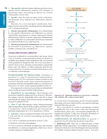
              The sequence in evolution of granuloma is schematically
           shown in Fig. 6.18 and is briefly outlined below:
           1. Engulfment by macrophages.  Macrophages and
           monocytes engulf the antigen and try to destroy it. But since  Figure 6.18  Mechanism of evolution of a granuloma (IL=interleukin;
           the antigen is poorly degradable, these cells fail to digest  IFN= interferon; TNF = tumour necrosis factor).
           and degrade the antigen, and instead undergo morphologic
           changes to epithelioid cells.                       iv) Growth factors (transforming growth factor-β, platelet-
                                                               derived growth factor) elaborated by activated macrophages
           2. CD4+ T cells. Macrophages, being antigen-presenting  stimulate fibroblast growth.
           cells, having failed to deal with the antigen, present it to CD4+  Thus, a granuloma is formed of macrophages modified
           T lymphocytes. These lymphocytes get activated and  as epithelioid cells in the centre, with some interspersed
           elaborate lymphokines (IL-1, IL-2, interferon-γ, TNF-α).  multinucleate giant cells, surrounded peripherally by
           3. Cytokines. Various cytokines formed by activated CD4+  lymphocytes (mainly T cells), and  healing by fibroblasts or
           T cells and also by activated macrophages perform the  collagen depending upon the age of granuloma.
           following roles:                                    COMPOSITION OF GRANULOMA.  In general, a
           i) IL-1 and IL-2 stimulate proliferation of more T cells.  granuloma has the following structural composition:
           ii) Interferon-γ activates macrophages.             1. Epithelioid cells. These are so called because of their
           iii) TNF-α promotes fibroblast proliferation and activates  epithelial cell-like appearance, are modified macrophages/
           endothelium to secrete prostaglandins which have role in  histiocytes which are somewhat elongated, having vesicular
           vascular response in inflammation.                  and lightly-staining slipper-shaped nucleus, and pale-

