


Page 181 - Textbook of Pathology, 6th Edition
P. 181
HEALING                          165
                                                               Healing is the body response to injury in an attempt to restore
                                                               normal structure and function. Healing involves 2 distinct
                                                               processes:
                                                                  Regeneration when healing takes place by proliferation of  CHAPTER 6
                                                               parenchymal cells and usually results in complete restoration
                                                               of the original tissues.
                                                                  Repair when healing takes place by proliferation of
                                                               connective tissue elements resulting in fibrosis and scarring.
                                                                  At times, both the processes take place simultaneously.

                                                               REGENERATION
                                                               Some parenchymal cells are short-lived while others have a
                                                               longer lifespan. In order to maintain proper structure of
                                                               tissues, these cells are under the constant regulatory control
                                                               of their cell cycle. These include growth factors such as:  Inflammation and Healing
                                                               epidermal growth factor, fibroblast growth factor, platelet-
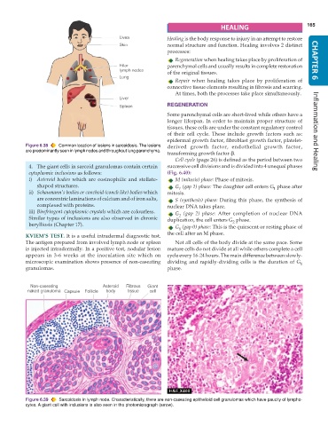
           Figure 6.38  Common location of lesions in sarcoidosis. The lesions  derived growth factor, endothelial growth factor,
           are predominantly seen in lymph nodes and throughout lung parenchyma.
                                                               transforming growth factor-β.
                                                                  Cell cycle (page 26) is defined as the period between two
            4. The giant cells in sarcoid granulomas contain certain  successive cell divisions and is divided into 4 unequal phases
            cytoplasmic inclusions as follows:                 (Fig. 6.40):
            i) Asteroid bodies which are eosinophilic and stellate-  M (mitosis) phase: Phase of mitosis.
               shaped structures.                                 G  (gap 1) phase: The daughter cell enters G  phase after
                                                                    1
            ii) Schaumann’s bodies or conchoid (conch like) bodies which  mitosis.                     1
               are concentric laminations of calcium and of iron salts,  S (synthesis) phase: During this phase, the synthesis of
               complexed with proteins.                        nuclear DNA takes place.
            iii) Birefringent cytoplasmic crystals which are colourless.  G  (gap 2) phase: After completion of nuclear DNA
                                                                    2
            Similar types of inclusions are also observed in chronic  duplication, the cell enters G  phase.
            berylliosis (Chapter 17).                                                   2
                                                                  G  (gap 0) phase: This is the quiescent or resting phase of
                                                                    0
                                                               the cell after an M phase.
           KVIEM’S TEST. It is a useful intradermal diagnostic test.
           The antigen prepared from involved lymph node or spleen  Not all cells of the body divide at the same pace. Some
           is injected intradermally. In a positive test, nodular lesion  mature cells do not divide at all while others complete a cell
           appears in 3-6 weeks at the inoculation site which on  cycle every 16-24 hours. The main difference between slowly-
           microscopic examination shows presence of non-caseating  dividing and rapidly-dividing cells is the duration of G 1
           granulomas.                                         phase.


           Figure 6.39  Sarcoidosis in lymph node. Characteristically, there are non-caseating epithelioid cell granulomas which have paucity of lympho-
           cytes. A giant cell with inclusions is also seen in the photomicrograph (arrow).

