


Page 182 - Textbook of Pathology, 6th Edition
P. 182
166


     SECTION I


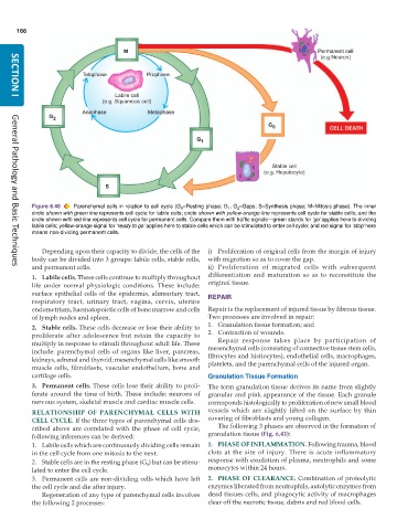
           Figure 6.40  Parenchymal cells in relation to cell cycle (G –Resting phase; G , G –Gaps; S–Synthesis phase; M–Mitosis phase). The inner
                                                                     1
                                                       0
                                                                        2
           circle shown with green line represents cell cycle for labile cells; circle shown with yellow-orange line represents cell cycle for stable cells; and the
           circle shown with red line represents cell cycle for permanent cells. Compare them with traffic signals—green stands for ‘go’ applies here to dividing
           labile cells; yellow-orange signal for ‘ready to go’ applies here to stable cells which can be stimulated to enter cell cycle; and red signal for ‘stop’ here
           means non-dividing permanent cells.
              Depending upon their capacity to divide, the cells of the  i) Proliferation of original cells from the margin of injury
           body can be divided into 3 groups: labile cells, stable cells,  with migration so as to cover the gap.
           and permanent cells.                                ii) Proliferation of migrated cells with subsequent
     General Pathology and Basic Techniques
           1. Labile cells. These cells continue to multiply throughout  differentiation and maturation so as to reconstitute the
           life under normal physiologic conditions. These include:  original tissue.
           surface epithelial cells of the epidermis, alimentary tract,  REPAIR
           respiratory tract, urinary tract, vagina, cervix, uterine
           endometrium, haematopoietic cells of bone marrow and cells  Repair is the replacement of injured tissue by fibrous tissue.
           of lymph nodes and spleen.                          Two processes are involved in repair:
           2. Stable cells. These cells decrease or lose their ability to  1. Granulation tissue formation; and
           proliferate after adolescence but retain the capacity to  2. Contraction of wounds.
           multiply in response to stimuli throughout adult life. These  Repair response takes place by participation of
           include: parenchymal cells of organs like liver, pancreas,  mesenchymal cells (consisting of connective tissue stem cells,
           kidneys, adrenal and thyroid; mesenchymal cells like smooth  fibrocytes and histiocytes), endothelial cells, macrophages,
           muscle cells, fibroblasts, vascular endothelium, bone and  platelets, and the parenchymal cells of the injured organ.
           cartilage cells.                                    Granulation Tissue Formation
           3. Permanent cells. These cells lose their ability to proli-  The term granulation tissue derives its name from slightly
           ferate around the time of birth. These include: neurons of  granular and pink appearance of the tissue. Each granule
           nervous system, skeletal muscle and cardiac muscle cells.  corresponds histologically to proliferation of new small blood
           RELATIONSHIP OF PARENCHYMAL CELLS WITH              vessels which are slightly lifted on the surface by thin
           CELL CYCLE. If the three types of parenchymal cells des-  covering of fibroblasts and young collagen.
           cribed above are correlated with the phase of cell cycle,  The following 3 phases are observed in the formation of
           following inferences can be derived:                granulation tissue (Fig. 6.41):
           1. Labile cells which are continuously dividing cells remain  1. PHASE OF INFLAMMATION. Following trauma, blood
           in the cell cycle from one mitosis to the next.     clots at the site of injury. There is acute inflammatory
           2. Stable cells are in the resting phase (G ) but can be stimu-  response with exudation of plasma, neutrophils and some
                                             0
           lated to enter the cell cycle.                      monocytes within 24 hours.
           3. Permanent cells are non-dividing cells which have left  2. PHASE OF CLEARANCE. Combination of proteolytic
           the cell cycle and die after injury.                enzymes liberated from neutrophils, autolytic enzymes from
              Regeneration of any type of parenchymal cells involves  dead tissues cells, and phagocytic activity of macrophages
           the following 2 processes:                          clear off the necrotic tissue, debris and red blood cells.

