


Page 180 - Textbook of Pathology, 6th Edition
P. 180
164


     SECTION I


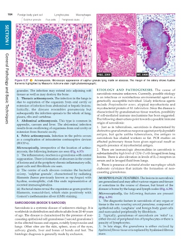
           Figure 6.37  Actinomycosis. Microscopic appearance of sulphur granule lying inside an abscess. The margin of the colony shows hyaline
           filaments highlighted by Masson’s trichrome stain (right photomicrograph).

            granules. The infection may extend into adjoining soft  ETIOLOGY AND PATHOGENESIS.  The cause of
            tissues as well as may destroy the bone.           sarcoidosis remains unknown. Currently, possible etiology
            2. Thoracic actinomycosis. The infection in the lungs is  is an infectious or noninfectious environmental agent in a
            due to aspiration of the organism from oral cavity or  genetically susceptible individual. Likely infectious agents
            extension of infection from abdominal or hepatic lesions.  include  Propionibacter acnes, atypical mycobacteria and
            Initially, the disease resembles pneumonia but     mycobacterial protein of M. tuberculosis. Since the disease is
            subsequently the infection spreads to the whole of lung,  characterised by granulomatous tissue reaction, possibility
            pleura, ribs and vertebrae.                        of cell-mediated immune mechanisms has been suggested.
            3. Abdominal actinomycosis. This type is common in  The following observations point towards a possible immune
            appendix, caecum and liver. The abdominal infection  origin of sarcoidosis:
     General Pathology and Basic Techniques
            results from swallowing of organisms from oral cavity or  1. Just as in tuberculosis, sarcoidosis is characterised by
            extension from thoracic cavity.                    distinctive granulomatous response against poorly degradable
            4. Pelvic actinomycosis. Infection in the pelvis occurs  antigen, but quite unlike tuberculosis, the antigen in
            as a complication of intrauterine contraceptive devices  sarcoidosis has eluded workers so far. PCR studies on
            (IUCD’s).                                          affected pulmonary tissue have given equivocal result as
                                                               regards presence of mycobacterial antigen.
            Microscopically, irrespective of the location of actino-  2. There are immunologic abnormalities in sarcoidosis is
            mycosis, the following features are seen (Fig. 6.37):  substantiated by high levels of  CD4+T cells lavaged from lung
            i) The inflammatory reaction is a granuloma with central  lesions. There is also elevation in levels of IL-2 receptors in
            suppuration. There is formation of abscesses in the centre  serum and in lavaged fluid from lungs.
            of lesions and at the periphery chronic inflammatory cells,
            giant cells and fibroblasts are seen.              3. There is presence of activated alveolar macrophages which
            ii) The centre of each abscess contains the bacterial  elaborate cytokines that initiate the formation of non-
            colony, ‘sulphur granule’, characterised by radiating  caseating granulomas.
            filaments (hence previously known as ray fungus) with  MORPHOLOGIC FEATURES. The lesions in sarcoidosis
            hyaline, eosinophilic, club-like ends representative of  are generalised and may affect various organs and tissues
            secreted immunoglobulins.                            at sometime in the course of disease, but brunt of the
            iii) Bacterial stains reveal the organisms as gram-positive  disease is borne by the lungs and lymph nodes (Fig. 6.38).
            filaments, nonacid-fast, which stain positively with  Microscopically, the following features are present
            Gomori’s methenamine silver (GMS) staining.          (Fig. 6.39):
                                                                 1. The diagnostic feature in sarcoidosis of any organ or
           SARCOIDOSIS (BOECK’S SARCOID)
                                                                 tissue is the non-caseating sarcoid granuloma, composed of
           Sarcoidosis is a systemic disease of unknown etiology. It is  epithelioid cells, Langhans’ and foreign body giant cells
           worldwide in distribution and affects adults from 20-40 years  and surrounded peripherally by fibroblasts.
           of age. The disease is characterised by the presence of non-  2. Typically, granulomas of sarcoidosis are ‘naked’ i.e.
           caseating epithelioid cell granulomas (‘sarcoid granuloma’)  either devoid of peripheral rim of lymphocytes or there is
           in the affected tissues and organs, notably lymph nodes and  paucity of lymphocytes.
           lungs. Other sites are the skin, spleen, uvea of the eyes,  3. In late stage, the granuloma is either  enclosed by
           salivary glands, liver and bones of hands and feet. The  hyalinised fibrous tissue or is replaced by hyalinised fibrous
           histologic diagnosis is generally made by exclusion.  mass.

