


Page 318 - Textbook of Pathology, 6th Edition
P. 318
302
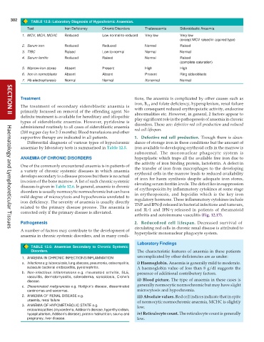
TABLE 12.5: Laboratory Diagnosis of Hypochromic Anaemias.
Test Iron Deficiency Chronic Disorders Thalassaemia Sideroblastic Anaemia
1. MCV, MCH, MCHC Reduced Low normal-to-reduced Very low Very low
(except MCV raised in aquired type)
2. Serum iron Reduced Reduced Normal Raised
3. TIBC Raised Low-to-normal Normal Normal
4. Serum ferritin Reduced Raised Normal Raised
(complete saturation)
5. Marrow-iron stores Absent Present High High
6. Iron in normoblasts Absent Absent Present Ring sideroblasts
7. Hb electrophoresis Normal Normal Abnormal Normal
Treatment tions, the anaemia is complicated by other causes such as
iron, B and folate deficiency, hypersplenism, renal failure
12
The treatment of secondary sideroblastic anaemia is with consequent reduced erythropoietic activity, endocrine
primarily focussed on removal of the offending agent. No abnormalities etc. However, in general, 2 factors appear to
definite treatment is available for hereditary and idiopathic play significant role in the pathogenesis of anaemia in chronic
SECTION II
types of sideroblastic anaemias. However, pyridoxine is
administered routinely to all cases of sideroblastic anaemia disorders. These are: defective red cell production and reduced
(200 mg per day for 2-3 months). Blood transfusions and other red cell lifespan.
supportive therapy are indicated in all patients. 1. Defective red cell production. Though there is abun-
Differential diagnosis of various types of hypochromic dance of storage iron in these conditions but the amount of
anaemias by laboratory tests is summarised in Table 12.5. iron available to developing erythroid cells in the marrow is
subnormal. The mononuclear phagocyte system is
ANAEMIA OF CHRONIC DISORDERS hyperplastic which traps all the available free iron due to
the activity of iron binding protein, lactoferrin. A defect in
One of the commonly encountered anaemia is in patients of the transfer of iron from macrophages to the developing
a variety of chronic systemic diseases in which anaemia erythroid cells in the marrow leads to reduced availability
develops secondary to a disease process but there is no actual of iron for haem synthesis despite adequate iron stores,
invasion of the bone marrow. A list of such chronic systemic elevating serum ferritin levels. The defect lies in suppression
diseases is given in Table 12.6. In general, anaemia in chronic of erythropoieitn by inflammatory cytokines at some stage
disorders is usually normocytic normochromic but can have in erythropoiesis, and hepcidin which is the key iron
mild degree of microcytosis and hypochromia unrelated to regulatory hormone. These inflammatory cytokines include
iron deficiency. The severity of anaemia is usually directly TNF and IFN-β released in bacterial infections and tumours,
related to the primary disease process. The anaemia is and IL-1 and IFN-γ released in patients of rheumatoid
corrected only if the primary disease is alleviated.
arthritis and autoimmune vasculitis (Fig. 12.17).
Pathogenesis 2. Reduced red cell lifespan. Decreased survival of
circulating red cells in chronic renal disease is attributed to
A number of factors may contribute to the development of hyperplastic mononuclear phagocyte system.
Haematology and Lymphoreticular Tissues
anaemia in chronic systemic disorders, and in many condi-
Laboratory Findings
TABLE 12.6: Anaemias Secondary to Chronic Systemic
Disorders. The characteristic features of anaemia in these patients
1. ANAEMIA IN CHRONIC INFECTIONS/INFLAMMATION uncomplicated by other deficiencies are as under:
a. Infections e.g. tuberculosis, lung abscess, pneumonia, osteomyelitis, i) Haemoglobin. Anaemia is generally mild to moderate.
subacute bacterial endocarditis, pyelonephritis. A haemoglobin value of less than 8 g/dl suggests the
b. Non-infectious inflammations e.g. rheumatoid arthritis, SLE, presence of additional contributory factors.
vasculitis, dermatomyositis, scleroderma, sarcoidosis, Crohn’s
disease. ii) Blood picture. The type of anaemia in these cases is
c. Disseminated malignancies e.g. Hodgkin’s disease, disseminated generally normocytic normochromic but may have slight
carcinomas and sarcomas. microcytosis and hypochromia.
2. ANAEMIA OF RENAL DISEASE e.g. iii) Absolute values. Red cell indices indicate that in spite
uraemia, renal failure of normocytic normochromic anaemia, MCHC is slightly
3. ANAEMIA OF HYPOMETABOLIC STATE e.g. low.
endocrinopathies (myxoedema, Addison's disease, hyperthyroidism,
hypopituitarism, Addison’s disease), protein malnutrition, scurvy and iv) Reticulocyte count. The reticulocyte count is generally
pregnancy, liver disease. low.

