


Page 335 - Textbook of Pathology, 6th Edition
P. 335
2.Mechanism of sickling: During deoxygenation, the red                                                   319
           cells containing HbS change from biconcave disc shape to
           an elongated crescent-shaped or sickle-shaped cell. This
           process termed sickling occurs both within the intact red cells
           and in vitro in free solution. The mechanism responsible for
           sickling upon deoxygenation of HbS-containing red cells is
           the polymerisation of deoxygenated HbS which aggregates
           to form elongated rod-like polymers. These elongated fibres
           align and distort the red cell into classic sickle shape.
           3. Reversible-irreversible sickling: The oxygen-dependent
           sickling process is usually reversible. However, damage to
           red cell membrane leads to formation of irreversibly sickled
           red cells even after they are exposed to normal oxygen
           tension.
           4.Factors determining rate of sickling: Following factors
           determine the rate at which the polymerisation of HbS and                                                  CHAPTER 12
           consequent sickling take place:
           i) Presence of non-HbS haemoglobins: The red cells in patients
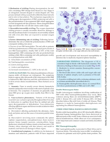
           of SS have predominance of HbS and a small part consists of  Figure 12.29  Sickle cell anaemia. PBF shows crescent shaped
           non-HbS haemoglobins, chiefly HbF (2-20% of the total  elongated red blood cells, a few target cells and a few erythroblasts.
           haemoglobin). HbF-containing red cells are protected from
           sickling while HbA-containing red cells participate readily  growth and development and increased susceptibility to
           in co-polymerisation with HbS.                      infection due to markedly impaired splenic function.
           ii) Intracellular concentration of HbS.
                                                                 LABORATORY FINDINGS.  The diagnosis of SS is
           iii) Total haemoglobin concentration.
                                                                 considered high in blacks with haemolytic anaemia. The
           iv) Extent of deoxygenation.
                                                                 laboratory findings in these cases are as under (Fig. 12.29):
           v) Acidosis and dehydration.
                                                                 1. Moderate to severe anaemia (haemoglobin concen-
           vi) Increased concentration of 2, 3-BPG in the red cells.  tration 6-9 g/dl).
           CLINICAL FEATURES. The clinical manifestations of homo-  2. The blood film shows sickle cells and target cells and
           zygous sickle cell disease are widespread. The symptoms  features of splenic atrophy such as presence of Howell-
           begin to appear after 6th month of life when most of the HbF  Jolly bodies.
           is replaced by HbS. Infection and folic acid deficiency result  3. A positive sickling test with a reducing substance such
           in more severe clinical manifestations. These features are as  as sodium metabisulfite (described above).
           under:                                                4. Haemoglobin electrophoresis shows no normal HbA
           1. Anaemia. There is usually severe chronic haemolytic  but shows predominance of HbS and 2-20% HbF.
           anaemia (primarily extravascular) with onset of aplastic crisis  Double Heterozygous States                Introduction to Haematopoietic System and Disorders of Erythroid Series
           in between. The symptoms of anaemia are generally mild
           since HbS gives up oxygen more readily than HbA to the  Double heterozygous conditions involving combination of
           tissues.                                            HbS with other haemoglobinopathies may occur. Most
                                                                                                            S thal
           2. Vaso-occlusive  phenomena.  Patients of SS develop  common among these are sickle-β-thalassaemia (β β  ),
           recurrent vaso-occlusive episodes throughout their lives due  sickle C disease (SC), and sickle D disease (SD). All these
           to obstruction to capillary blood flow by sickled red cells  disorders behave like mild form of sickle cell disease. Their
           upon deoxygenation or dehydration. Vaso-obstruction  diagnosis is made by haemoglobin electrophoresis and
           affecting different organs and tissues results in infarcts which  separating the different haemoglobins.
           may be of 2 types:                                  OTHER STRUCTURAL HAEMOGLOBINOPATHIES
           i) Microinfarcts affecting particularly the abdomen, chest,  Besides sickle haemoglobin, about 400 structurally different
           back and joints and are the cause of recurrent painful crises  abnormal human haemoglobins have been discovered in
           in SS.                                              different parts of the world. Some of them are associated with
           ii) Macroinfarcts involving most commonly the spleen  clinical manifestations, while others are of no consequence.
           (splenic sequestration, autosplenectomy), bone marrow  A few important and common variants are briefly described
           (pains), bones (aseptic necrosis, osteomyelitis), lungs  below:
           (pulmonary infections), kidneys (renal cortical necrosis), CNS
           (stroke), retina (damage) and skin (ulcers), and result in  HbC Haemoglobinopathy
           anatomic and functional damage to these organs.     HbC haemoglobinopathy is prevalent in West Africa and in
           3. Constitutional symptoms. In addition to the features of  American blacks. The molecular lesion in HbC is substitution
           anaemia and infarction, patients with SS have impaired  of lysine for glutamic acid at β-6 globin chain position. The

