


Page 343 - Textbook of Pathology, 6th Edition
P. 343
Disorders of Platelets,                                            327

            Chapter 13
            Chapter 13                         Bleeding Disorders and

                                           Basic Transfusion Medicine


                         THROMBOPOIESIS                        granules. Platelets are formed from pseudopods of
                                                               megakaryocyte cytoplasm which get detached into the blood
           As outlined in previous chapter and illustrated in Fig. 12.3,  stream. Each megakaryocyte may form up to 4000 platelets.
           the trilineage myeloid stem cells in the bone marrow  The formation of platelets from the stem cell takes about
           differentiate into erythroid progenitor, granulocyte-  10 days.
           monocyte progenitor, and megakaryocyte progenitor cells.  PLATELETS.  Platelets are small (1-4  μm in diameter),
           Platelets are formed in the bone marrow by a process of  discoid, non-nucleate structures containing red-purple  CHAPTER 13
           fragmentation of the cytoplasm of megakaryocytes. Platelet  granules. The normal platelet count ranges from 150,000-
           production is under the control of thrombopoietin, the nature  400,000/μl and the lifespan of platelets is 7-10 days. About
           and origin of which are not yet established. The stages in  70% of platelets are in circulation while remaining 30% lie
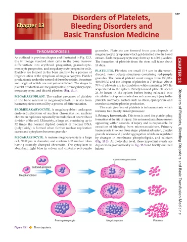
           platelet production are: megakaryoblast, promegakaryocyte,  sequestered in the spleen. Newly-formed platelets spend
           megakaryocyte, and discoid platelets (Fig. 13.1).
                                                               24-36 hours in the spleen before being released into
           MEGAKARYOBLAST. The earliest precursor of platelets  circulation but splenic stasis does not cause any injury to the
           in the bone marrow is megakaryoblast. It arises from  platelets normally. Factors such as stress, epinephrine and
           haematopoietic stem cell by a process of differentiation.  exercise stimulate platelet production.
                                                                  The main functions of platelets is in haemostasis which
           PROMEGAKARYOCYTE.  A megakaryoblast undergoes       includes two closely linked processes:
           endo-reduplication of nuclear chromatin i.e. nuclear
           chromatin replicates repeatedly in multiples of two without  1. Primary haemostasis. This term is used for platelet plug
           division of the cell. Ultimately, a large cell containing up to  formation at the site of injury. It is an immediate phenomenon
           32 times the normal diploid content of nuclear DNA  appearing within seconds of injury and is responsible for
           (polyploidy) is formed when further nuclear replication  cessation of bleeding from microvasculature. Primary
           ceases and cytoplasm becomes granular.              haemostasis involves three steps: platelet adhesion, platelet
                                                               granule release and platelet aggregation which are regulated
           MEGAKARYOCYTE. A mature megakaryocyte is a large    by changes in membrane phospholipids, and calcium
           cell, 30-90 μm in diameter, and contains 4-16 nuclear lobes  (Fig. 13.2). At molecular level, these important events are
           having coarsely clumped chromatin. The cytoplasm is  depicted diagrammatically in Fig. 13.3 and briefly outlined
           abundant, light blue in colour and contains red-purple  below:                                             Disorders of Platelets, Bleeding Disorders and Basic Transfusion Medicine


           Figure 13.1  Thrombopoiesis.

