


Page 438 - Textbook of Pathology, 6th Edition
P. 438
422


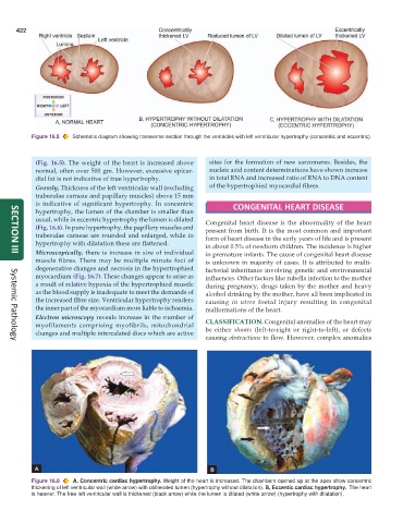
           Figure 16.5  Schematic diagram showing transverse section through the ventricles with left ventricular hypertrophy (concentric and eccentric).


            (Fig. 16.5). The weight of the heart is increased above  sites for the formation of new sarcomeres. Besides, the
            normal, often over 500 gm. However, excessive epicar-  nucleic acid content determinations have shown increase
            dial fat is not indicative of true hypertrophy.      in total RNA and increased ratio of RNA to DNA content
            Grossly, Thickness of the left ventricular wall (excluding  of the hypertrophied myocardial fibres.
            trabeculae carneae and papillary muscles) above 15 mm
            is indicative of significant hypertrophy. In concentric     CONGENITAL HEART DISEASE
            hypertrophy, the lumen of the chamber is smaller than
            usual, while in eccentric hypertrophy the lumen is dilated  Congenital heart disease is the abnormality of the heart
            (Fig. 16.6). In pure hypertrophy, the papillary muscles and  present from birth. It is the most common and important
            trabeculae carneae are rounded and enlarged, while in  form of heart disease in the early years of life and is present
            hypertrophy with dilatation these are flattened.   in about 0.5% of newborn children. The incidence is higher
            Microscopically, there is increase in size of individual  in premature infants. The cause of congenital heart disease
     SECTION III
            muscle fibres. There may be multiple minute foci of  is unknown in majority of cases. It is attributed to multi-
            degenerative changes and necrosis in the hypertrophied  factorial inheritance involving genetic and environmental
            myocardium (Fig. 16.7). These changes appear to arise as  influences. Other factors like rubella infection to the mother
            a result of relative hypoxia of the hypertrophied muscle  during pregnancy, drugs taken by the mother and heavy
            as the blood supply is inadequate to meet the demands of  alcohol drinking by the mother, have all been implicated in
            the increased fibre size. Ventricular hypertrophy renders  causing  in utero foetal injury resulting in congenital
            the inner part of the myocardium more liable to ischaemia.  malformations of the heart.
            Electron microscopy reveals increase in the number of  CLASSIFICATION. Congenital anomalies of the heart may
            myofilaments comprising myofibrils, mitochondrial  be either  shunts (left-to-right or right-to-left), or defects
            changes and multiple intercalated discs which are active
                                                               causing obstructions to flow. However, complex anomalies
     Systemic Pathology


           Figure 16.6  A, Concentric cardiac hypertrophy. Weight of the heart is increased. The chambers opened up at the apex show concentric
           thickening of left ventricular wall (white arrow) with obliterated lumen (hypertrophy without dilatation). B, Eccentic cardiac hypertrophy.  The heart
           is heavier. The free left ventricular wall is thickened (black arrow) while the lumen is dilated (white arrow) (hypertrophy with dilatation).

