


Page 437 - Textbook of Pathology, 6th Edition
P. 437
421


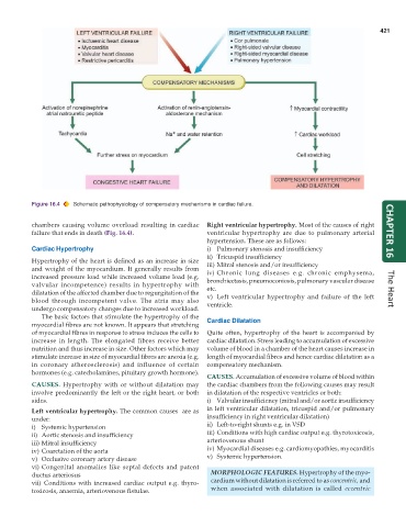
           Figure 16.4  Schematic pathophysiology of compensatory mechanisms in cardiac failure.


           chambers causing volume overload resulting in cardiac  Right ventricular hypertrophy. Most of the causes of right
           failure that ends in death (Fig. 16.4).             ventricular hypertrophy are due to pulmonary arterial  CHAPTER 16
                                                               hypertension. These are as follows:
           Cardiac Hypertrophy                                 i) Pulmonary stenosis and insufficiency
                                                               ii) Tricuspid insufficiency
           Hypertrophy of the heart is defined as an increase in size  iii) Mitral stenosis and/or insufficiency
           and weight of the myocardium. It generally results from  iv) Chronic lung diseases e.g. chronic emphysema,
           increased pressure load while increased volume load (e.g.  bronchiectasis, pneumoconiosis, pulmonary vascular disease
           valvular incompetence) results in hypertrophy with  etc.
           dilatation of the affected chamber due to regurgitation of the  v) Left ventricular hypertrophy and failure of the left  The Heart
           blood through incompetent valve. The atria may also  ventricle.
           undergo compensatory changes due to increased workload.
              The basic factors that stimulate the hypertrophy of the
           myocardial fibres are not known. It appears that stretching  Cardiac Dilatation
           of myocardial fibres in response to stress induces the cells to  Quite often, hypertrophy of the heart is accompanied by
           increase in length. The elongated fibres receive better  cardiac dilatation. Stress leading to accumulation of excessive
           nutrition and thus increase in size. Other factors which may  volume of blood in a chamber of the heart causes increase in
           stimulate increase in size of myocardial fibres are anoxia (e.g.  length of myocardial fibres and hence cardiac dilatation as a
           in coronary atherosclerosis) and influence of certain  compensatory mechanism.
           hormones (e.g. catecholamines, pituitary growth hormone).
                                                               CAUSES. Accumulation of excessive volume of blood within
           CAUSES.  Hypertrophy with or without dilatation may  the cardiac chambers from the following causes may result
           involve predominantly the left or the right heart, or both  in dilatation of the respective ventricles or both:
           sides.                                              i) Valvular insufficiency (mitral and/or aortic insufficiency
           Left ventricular hypertrophy. The common causes  are as  in left ventricular dilatation, tricuspid and/or pulmonary
           under:                                              insufficiency in right ventricular dilatation)
           i) Systemic hypertension                            ii) Left-to-right shunts e.g. in VSD
           ii) Aortic stenosis and insufficiency               iii) Conditions with high cardiac output e.g. thyrotoxicosis,
           iii) Mitral insufficiency                           arteriovenous shunt
           iv) Coarctation of the aorta                        iv) Myocardial diseases e.g. cardiomyopathies, myocarditis
           v) Occlusive coronary artery disease                v) Systemic hypertension.
           vi) Congenital anomalies like septal defects and patent
           ductus arteriosus                                     MORPHOLOGIC FEATURES. Hypertrophy of the myo-
           vii) Conditions with increased cardiac output e.g. thyro-  cardium without dilatation is referred to as concentric, and
           toxicosis, anaemia, arteriovenous fistulae.           when associated with dilatation is called  eccentric

