


Page 439 - Textbook of Pathology, 6th Edition
P. 439
II. SHUNTS (CYANOTIC CONGENITAL HEART DISEASE) 423
A shunt may be left-to-right side or right-to-left side of the
circulation.
A. Left-to-Right Shunts
(Acyanotic or Late Cyanotic Group)
In conditions where there is shunting of blood from left-to-
right side of the heart, there is volume overload on the right
heart producing pulmonary hypertension and right
ventricular hypertrophy. At a later stage, the pressure on
the right side is higher than on the left side creating late
cyanotic heart disease. The important conditions included
in this category are described below:
VENTRICULAR SEPTAL DEFECT (VSD). VSD is the most
common congenital anomaly of the heart and comprises
about 30% of all congenital heart diseases. The condition is
recognised early in life. The smaller defects often close
spontaneously, while larger defects remain patent and
produce significant effects.
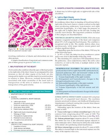
Figure 16.7 Cardiac hypertrophy. Individual myocardial fibres are
thick with prominent vesicular nuclei. Depending upon the location of the defect, VSD may be
of the following types:
1. In 90% of cases, the defect involves membranous septum
involving combinations of shunts and obstructions are also and is very close to the bundle of His (Fig. 16.8).
often present. 2. The remaining 10% cases have VSD immediately below
A simple classification of important and common exam- the pulmonary valve (subpulmonic), below the aortic valve
ples of these groups is given in Table 16.1. (subaortic), or exist in the form of multiple defects in the CHAPTER 16
muscular septum.
I. MALPOSITIONS OF THE HEART
MORPHOLOGIC FEATURES. The effects of VSD are
Dextrocardia is the condition when the apex of the heart points produced due to left-to-right shunt at the ventricular level,
to the right side of the chest. It may be accompanied by situs increased pulmonary flow and increased volume in the
inversus so that all other organs of the body are also left side of the heart. These effects are as under:
transposed in similar way and thus heart is in normal position i) Volume hypertrophy of the right ventricle. The Heart
in relation to them. However, isolated dextrocardia is ii) Enlargement and haemodynamic changes in the
associated with major anomalies of the heart such as tricuspid and pulmonary valves.
transposition of the atria in relation to ventricles or iii) Endocardial hypertrophy of the right ventricle.
transposition of the great arteries. iv) Pressure hypertrophy of the right atrium.
v) Volume hypertrophy of the left atrium and left
ventricle.
TABLE 16.1: Classification of Congenital Heart Diseases. vi) Enlargement and haemodynamic changes in the mitral
I. MALPOSITIONS OF THE HEART and aortic valves.
II. SHUNTS ATRIAL SEPTAL DEFECT (ASD). Isolated ASD comprises
(CYANOTIC CONGENITAL HEART DISEASE) about 10% of congenital heart diseases. The condition
A. Left-to-right shunts remains unnoticed in infancy and childhood till pulmonary
(Acyanotic or late cyanotic group) hypertension is induced causing late cyanotic heart disease
1. Ventricular septal defect (VSD) (25-30%) and right-sided heart failure.
2. Atrial septal defect (ASD) (10-15%) Depending upon the location of the defect, there are
3. Patent ductus arteriosus (PDA) (10-20%) 3 types of ASD:
B. Right-to-left shunts (Cyanotic group) i) Fossa ovalis type or ostium secundum type is the most
1. Tetralogy of Fallot (6-15%) common form comprising about 90% cases of ASD. The
2. Transposition of great arteries (4-10%) defect is situated in the region of the fossa ovalis (Fig. 16.9).
3. Persistent truncus arteriosus (2%) ii) Ostium primum type comprises about 5% cases of ASD.
4. Tricuspid atresia and stenosis (1%) The defect lies low in the interatrial septum adjacent to
III. OBSTRUCTIONS atrioventricular valves. There may be cleft in the aortic leaflet
(OBSTRUCTIVE CONGENITAL HEART DISEASE) of the mitral valve producing mitral insufficiency.
1. Coarctation of aorta (5-7%) iii) Sinus venosus type accounts for about 5% cases of ASD.
2. Aortic stenosis and atresia (4-6%) The defect is located high in the interatrial septum near the
3. Pulmonary stenosis and atresia (5-7%)
entry of the superior vena cava.

