


Page 441 - Textbook of Pathology, 6th Edition
P. 441
The severity of the clinical manifestations is related to  425
                                                               two factors: extent of pulmonary stenosis and the size of VSD.
                                                               Accordingly, there are two forms of tetralogy: cyanotic and
                                                               acyanotic:
                                                               a) Cyanotic tetralogy: Pulmonary stenosis is greater and the
                                                               VSD is mild so that there is more resistance to the outflow of
                                                               blood from right ventricle resulting in right-to-left shunt at
                                                               the ventricular level and cyanosis. The effects on the heart
                                                               are as follows:
                                                               i) Pressure hypertrophy of the right atrium and right
                                                               ventricle.
                                                               ii) Smaller and abnormal tricuspid valve.
                                                               iii) Smaller left atrium and left ventricle.
                                                               iv) Enlarged aortic orifice.
                                                               b) Acyanotic tetralogy:  The VSD is larger and pulmonary
                                                               stenosis is mild so that there is mainly left-to-right shunt with
                                                               increased pulmonary flow and increased volume in the left
                                                               heart but no cyanosis. The effects on the heart are as under:
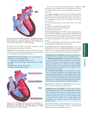
           Figure 16.10  Patent ductus arteriosus, a schematic representation  i) Pressure hypertrophy of the right ventricle and right
           (LA = Left atrium; LV = Left ventricle; PT = Pulmonary trunk; PV =  atrium.
           Pulmonary vein, AO = Aorta; RA = Right atrium; RV = Right ventricle;
           SVC = Superior vena cava; IVC = Inferior vena cava).  ii) Volume hypertrophy of the left atrium and left ventricle.
                                                               iii) Enlargement of mitral and aortic orifices.

           described here since there is functional shunting of blood  TRANSPOSITION OF GREAT ARTERIES.  The term
           from one to the other side of circulation.          transposition is used for complex malformations as regards
           TETRALOGY OF FALLOT. Tetralogy of Fallot is the most  position of the aorta, pulmonary trunk, atrioventricular  CHAPTER 16
           common cyanotic congenital heart disease, found in about  orifices and the position of atria in relation to ventricles.
           10% of children with anomalies of the heart.
                                                                 MORPHOLOGIC FEATURES. There are several forms of
            MORPHOLOGIC FEATURES. The four features of           transpositions. The common ones are described below:
            tetralogy are as under (Fig. 16.11):                 i) Regular transposition is the most common type. In this,
            i) Ventricular septal defect (VSD) (‘shunt’).        the aorta which is normally situated to the right and
            ii) Displacement of the aorta to right so that it overrides  posterior with respect to the pulmonary trunk, is instead  The Heart
            the VSD.                                             displaced anteriorly and to right. In regular complete
            iii) Pulmonary stenosis (‘obstruction’).             transposition, the aorta emerges from the right ventricle
            iv) Right ventricular hypertrophy.                   and the pulmonary trunk from the left ventricle so that
                                                                 there is cyanosis from birth.
                                                                 ii) Corrected transposition is an uncommon anomaly.
                                                                 There is complete transposition of the great arteries with
                                                                 aorta arising from the right ventricle and the pulmonary
                                                                 trunk from the left ventricle, as well as transposition of the
                                                                 great veins so that the pulmonary veins enter the right
                                                                 atrium and the systemic veins drain into the left atrium.
                                                                 This results in a physiologically corrected circulation.
                                                               PERSISTENT TRUNCUS ARTERIOSUS.  Persistent
                                                               truncus arteriosus (PTA) is a rare anomaly.
                                                                 MORPHOLOGIC FEATURES.  In PTA, the arch that
                                                                 normally separates the aorta from the pulmonary artery
                                                                 fails to develop. This results in a single large common
                                                                 vessel receiving blood from the right as well as left
                                                                 ventricle. The orifice may have 3 to 6 cusps. There is often
                                                                 an associated VSD. There is left-to-right shunt and
                                                                 frequently early systemic cyanosis. The prognosis is
                                                                 generally poor.
           Figure 16.11  Tetralogy of Fallot, a schematic representation (LA =  TRICUSPID ATRESIA AND STENOSIS. Tricuspid atresia
           Left atrium; LV = Left ventricle; PT = Pulmonary trunk; PV = Pulmonary
           vein; AO = Aorta; RA = Right atrium; RV = Right ventricle; SVC = Supe-  and stenosis are rare anomalies. There is often associated
           rior vena cava; IVC = Inferior vena cava).          pulmonary stenosis or pulmonary atresia.

