


Page 442 - Textbook of Pathology, 6th Edition
P. 442
426
            MORPHOLOGIC FEATURES. In tricuspid atresia, there    pressure in the lower extremities and effects of arterial
            is absence of tricuspid orifice and instead there is a dimple  insufficiency such as claudication and coldness. In time,
            in the floor of the right atrium. In tricuspid stenosis, the  there is development of collateral circulation between pre-
            tricuspid ring is small and the valve cusps are malformed.  stenotic and post-stenotic arterial branches so that
            In both the conditions, there is often an interatrial defect  intercostal arteries are enlarged and palpable and may
            through which right-to-left shunt of blood takes place.  produce erosions on the inner surface of the ribs.
            Children are cyanotic since birth and live for a few weeks  ii) Preductal or infantile type: The manifestations are
            or months.                                           produced early in life. The narrowing is proximal to the
                                                                 ductus arteriosus which usually remains patent. The
           III. OBSTRUCTIONS                                     narrowing is generally gradual and involves larger
             (OBSTRUCTIVE CONGENITAL HEART DISEASE)
                                                                 segment of the proximal aorta. There is often associated
           Congenital obstruction to blood flow may result from  interatrial septal defect. Preductal coarctation results in
           obstruction in the aorta due to narrowing (coarctation of aorta),  right ventricular hypertrophy while the left ventricle is
           obstruction to outflow from the left ventricle (aortic stenosis  small. Cyanosis develops in the lower half of the body
           and atresia), and obstruction to outflow from the right  while the upper half remains unaffected since it is supp-
           ventricle (pulmonary stenosis and atresia).           lied by vessels originating proximal to the coarctation.
                                                                 Children with this defect have poor prognosis.
           COARCTATION OF AORTA.  The word ‘coarctation’
           means contracted or compressed. Coarctation of aorta is  AORTIC STENOSIS AND ATRESIA. The most common
           localised narrowing in any part of aorta, but the constriction  congenital anomaly of the aorta is bicuspid aortic valve which
           is more often just distal to ductus arteriosus (postductal or  does not have much functional significance but predisposes
           adult), or occasionally proximal to the ductus arteriosus  it to calcification (page 450). Congenital aortic atresia is rare
           (preductal or infantile type) in the region of transverse aorta:
                                                               and incompatible with survival. Aortic stenosis may be
            MORPHOLOGIC FEATURES. The two common forms of      acquired (e.g. in rheumatic heart disease, calcific aortic
            coarrctation of the aorta are as under:            stenosis) or congenital.
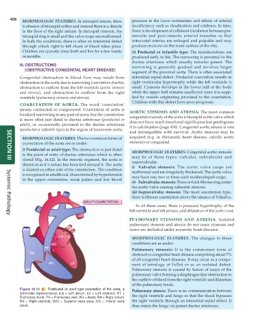
            i) Postductal or adult type: The obstruction is just distal  MORPHOLOGIC FEATURES. Congenital aortic stenosis
            to the point of entry of ductus arteriosus which is often  may be of three types: valvular, subvalvular and
            closed (Fig. 16.12). In the stenotic segment, the aorta is  supravalvular.
     SECTION III
            drawn in as if a suture has been tied around it. The aorta  i) Valvular stenosis: The aortic valve cusps are
            is dilated on either side of the constriction. The condition  malformed and are irregularly thickened. The aortic valve
            is recognised in adulthood, characterised by hypertension  may have one, two or three such maldeveloped cusps.
            in the upper extremities, weak pulses and low blood
                                                                 ii) Subvalvular stenosis: There is thick fibrous ring under
                                                                 the aortic valve causing subaortic stenosis.
                                                                 iii) Supravalvular stenosis: The most uncommon type,
                                                                 there is fibrous constriction above the sinuses of Valsalva.

                                                                  In all these cases, there is pressure hypertrophy of the
                                                               left ventricle and left atrium, and dilatation of the aortic root.
     Systemic Pathology
                                                               PULMONARY STENOSIS AND ATRESIA.  Isolated
                                                               pulmonary stenosis and atresia do not cause cyanosis and
                                                               hence are included under acyanotic heart diseases.

                                                                 MORPHOLOGIC FEATURES. The changes in these
                                                                 conditions are as under:
                                                                 Pulmonary stenosis: It is the commonest form of
                                                                 obstructive congenital heart disease comprising about 7%
                                                                 of all congenital heart diseases. It may occur as a compo-
                                                                 nent of tetralogy of Fallot or as an isolated defect.
                                                                 Pulmonary stenosis is caused by fusion of cusps of the
                                                                 pulmonary valve forming a diaphragm-like obstruction to
                                                                 the outflow of blood from the right ventricle and dilatation
                                                                 of the pulmonary trunk.
           Figure 16.12  Postductal or adult type coarctation of the aorta, a  Pulmonary atresia: There is no communication between
           schematic representation (LA = Left atrium; LV = Left ventricle; PT =  the right ventricle and lungs so that the blood bypasses
           Pulmonary trunk; PV = Pulmonary vein; AO = Aorta; RA = Right atrium;
           RV = Right ventricle; SVC = Superior vena cava; IVC = Inferior vena  the right ventricle through an interatrial septal defect. It
           cava).                                                then enters the lungs via patent ductus arteriosus.

