


Page 440 - Textbook of Pathology, 6th Edition
P. 440
424


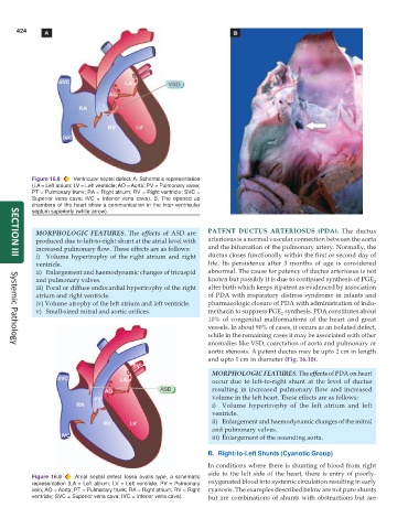
           Figure 16.8  Ventricular septal defect. A, Schematic representation
           (LA = Left atrium; LV = Left ventricle; AO = Aorta; PV = Pulmonary valve;
           PT = Pulmonary trunk; RA = Right atrium; RV = Right ventricle; SVC =
           Superior vena cava; IVC = Inferior vena cava). B, The opened up
           chambers of the heart show a communication in the inter-ventricular
           septum superiorly (white arrow).


            MORPHOLOGIC FEATURES. The effects of ASD are       PATENT DUCTUS ARTERIOSUS (PDA).  The ductus
            produced due to left-to-right shunt at the atrial level with  arteriosus is a normal vascular connection between the aorta
            increased pulmonary flow. These effects are as follows:  and the bifurcation of the pulmonary artery. Normally, the
            i) Volume hypertrophy of the right atrium and right  ductus closes functionally within the first or second day of
     SECTION III
            ventricle.                                         life. Its persistence after 3 months of age is considered
            ii) Enlargement and haemodynamic changes of tricuspid  abnormal. The cause for patency of ductus arteriosus is not
            and pulmonary valves.                              known but possibly it is due to continued synthesis of PGE 2
            iii) Focal or diffuse endocardial hypertrophy of the right  after birth which keeps it patent as evidenced by association
            atrium and right ventricle.                        of PDA with respiratory distress syndrome in infants and
            iv) Volume atrophy of the left atrium and left ventricle.  pharmacologic closure of PDA with administration of indo-
            v) Small-sized mitral and aortic orifices.         methacin to suppress PGE  synthesis. PDA constitutes about
                                                                                     2
                                                               10% of congenital malformations of the heart and great
                                                               vessels. In about 90% of cases, it occurs as an isolated defect,
                                                               while in the remaining cases it may be associated with other
                                                               anomalies like VSD, coarctation of aorta and pulmonary or
     Systemic Pathology
                                                               aortic stenosis. A patent ductus may be upto 2 cm in length
                                                               and upto 1 cm in diameter (Fig. 16.10).
                                                                 MORPHOLOGIC FEATURES. The effects of PDA on heart
                                                                 occur due to left-to-right shunt at the level of ductus
                                                                 resulting in increased pulmonary flow and increased
                                                                 volume in the left heart. These effects are as follows:
                                                                 i) Volume hypertrophy of the left atrium and left
                                                                 ventricle.
                                                                 ii) Enlargement and haemodynamic changes of the mitral
                                                                 and pulmonary valves.
                                                                 iii) Enlargement of the ascending aorta.

                                                               B. Right-to-Left Shunts (Cyanotic Group)
                                                               In conditions where there is shunting of blood from right
                                                               side to the left side of the heart, there is entry of poorly-
           Figure 16.9  Atrial septal defect fossa ovalis type, a schematic
           representation (LA = Left atrium; LV = Left ventricle; PV = Pulmonary  oxygenated blood into systemic circulation resulting in early
           vein; AO = Aorta; PT = Pulmonary trunk; RA = Right atrium; RV = Right  cyanosis. The examples described below are not pure shunts
           ventricle; SVC = Superior vena cava; IVC = Inferior vena cava).  but are combinations of shunts with obstructions but are

