


Page 492 - Textbook of Pathology, 6th Edition
P. 492
476


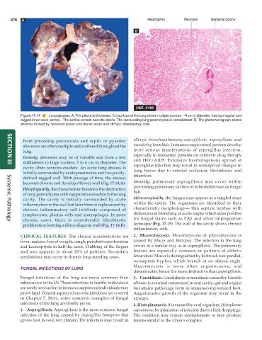
           Figure 17.14  Lung abscess. A, The pleura is thickened. Cut surface of the lung shows multiple cavities 1-4 cm in diameter, having irregular and
           ragged inner walls (arrow).  The lumina contain necrotic debris. The surrounding lung parenchyma is consolidated. B, The photomicrograph shows
           abscess formed by necrosed alveoli and dense acute and chronic inflammatory cells.


            from preceding pneumonia and septic or pyaemic     allergic bronchopulmonary aspergillosis, aspergilloma and
            abscesses are often multiple and scattered throughout the  necrotising bronchitis. Immunocompromised persons develop
            lung.                                              more serious manifestations of aspergillus infection,
                                                               especially in leukaemic patients on cytotoxic drug therapy
            Grossly, abscesses may be of variable size from a few  and HIV/AIDS. Extensive haematogenous spread of
            millimeters to large cavities, 5 to 6 cm in diameter. The  aspergillus infection may result in widespread changes in
     SECTION III
            cavity often contains exudate. An acute lung abscess is  lung tissue due to arterial occlusion, thrombosis and
            initially surrounded by acute pneumonia and has poorly-  infarction.
            defined ragged wall. With passage of time, the abscess
            becomes chronic and develops fibrous wall (Fig. 17.14,A).  Grossly, pulmonary aspergillosis may occur within
            Histologically, the characteristic feature is the destruction  preexisting pulmonary cavities or in bronchiectasis as fungal
                                                               ball.
            of lung parenchyma with suppurative exudate in the lung
            cavity. The cavity is initially surrounded by acute  Microscopically, the fungus may appear as a tangled mass
            inflammation in the wall but later there is replacement by  within the cavity. The organisms are identified by their
            chronic inflammatory cell infiltrate composed of   characteristic morphology— thin septate hyphae with
            lymphocytes, plasma cells and macrophages. In more  dichotomous branching at acute angles which stain positive
            chronic cases, there is considerable fibroblastic  for fungal stains such as PAS and silver impregnation
     Systemic Pathology
            proliferation forming a fibrocollagenic wall (Fig. 17.14,B).  technique (Fig. 17.15). The wall of the cavity shows chronic
                                                               inflammatory cells.
           CLINICAL FEATURES.  The clinical manifestations are  2. Mucormycosis. Mucormycosis or phycomycosis is
           fever, malaise, loss of weight, cough, purulent expectoration  caused by  Mucor and  Rhizopus. The infection in the lung
           and haemoptysis in half the cases. Clubbing of the fingers  occurs in a similar way as in aspergillosis. The pulmonary
           and toes appears in about 20% of patients. Secondary  lesions are especially common in patients of  diabetic
           amyloidosis may occur in chronic long-standing cases.  ketoacidosis. Mucor is distinguished by its broad, non-parallel,
                                                               nonseptate hyphae which branch at an obtuse angle.
                                                               Mucormycosis is more often angioinvasive, and
           FUNGAL INFECTIONS OF LUNG
                                                               disseminates; hence it is more destructive than aspergillosis.
           Fungal infections of the lung are more common than  3. Candidiasis. Candidiasis or moniliasis caused by Candida
           tuberculosis in the US. These infections in healthy individuals  albicans is a normal commensal in oral cavity, gut and vagina
           are rarely serious but in immunosuppressed individuals may  but attains pathologic form in immunocompromised host.
           prove fatal. General aspects of mycotic infections are covered  Angioinvasive growth of the organism may occur in the
           in Chapter 7. Here, some common examples of fungal  airways.
           infections of the lung are briefly given:
                                                               4. Histoplasmosis. It is caused by oval organism, Histoplasma
           1. Aspergillosis. Aspergillosis is the most common fungal  capsulatum, by inhalation of infected dust or bird droppings.
           infection of the lung caused by  Aspergillus fumigatus that  The condition may remain asymptomatic or may produce
           grows best in cool, wet climate. The infection may result in  lesions similar to the Ghon’s complex.

